

Introduction
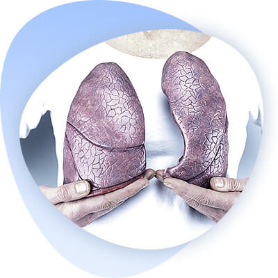
The larynx is an unpaired organ of complex structure; it is where the respiratory and digestive tracts intersect; it has protective, respiratory, and vocal functions.
Laryngeal cancer is a cancerous disease from elements of the non-keratinizing epithelium of the larynx. Laryngeal cancer accounts for 3% of all cancers. Among cancerous diseases of ENT organs, it is the most common (50-60%). In the structure of male cancer morbidity, laryngeal cancer ranks 5th. The incidence of laryngeal cancer in men is 7.39 cases per 100,000 people. Laryngeal cancer predominantly occurs in men 40-60 years old, comprising 80-95% of patients. The tumor is most often localized in the vestibular part of the larynx. Most patients are found to have stage III tumors (in 63% of cases). Advanced laryngeal cancer stage IV is detected in 16% of patients who seek medical help for the first time.

The vast majority of patients with laryngeal cancer have a long history of smoking, drinking alcohol, working in conditions of high dustiness and high temperature. Of particular importance is dust containing radioactive or harmful chemicals, and metal dust. In industrial cities, laryngeal cancer is 1.5-2 times more common than in rural areas.
Etiology
Contributing factors
Many harmful influences contribute to the occurrence of laryngeal cancer. The vast majority of patients can be noted in the anamnesis:
- A long period of smoking, alcohol consumption,
- work in conditions of excessive dustiness (dust containing radioactive or hazardous chemical substances, metal dust is of particular importance), high temperature etc.
The urban population falls ill with laryngeal cancer 1.5-2 times more often than the rural population. When explaining the incidence of cancer in a particular region, we have to analyze many factors: the degree of urbanization, geographic features, the predominant profile of industrial enterprises, local customs, and many others.
Precancerous diseases of the larynx
Precancerous diseases of the larynx include.
A. With a high incidence of oculosis (obligate):
- Dyskeratoses (leukoplakia, leukokeratosis);
- Pachydermia;
- Papilloma in adults. Men over 40 years of age are more often affected by these diseases.
B. With a low incidence of oselocation (facultative):
- Contact fibroma;
- Scar process after chronic specific infections (tuberculosis, syphilis, scleroma) and burns.
A. On examination, leukoplakia appears as a limited whitish-smoky spot, 2 to 10 mm in size, almost not elevated above the surface of the mucosa. The formations are more often single, but occasionally there are also multiple ones. Leukokeratosis is a keratinization of the mucous membrane. Leukokeratosis lesion looks like a grayish-white plaque with an uneven villous surface. The symptomatology of these diseases of the larynx is extremely poor. Patients note farting, dryness and a slight burning sensation in the throat, coughing, and occasional hoarseness. All patients, as a rule, - a chronic smoker. They attribute these sensations to the consequences of smoking, and only gradually the persistence of symptoms makes them see a doctor. Pachydermia - epidermoid outgrowths located near the vocal processes of the scapular cartilages or in the interscapular region (so-called laryngeal calluses) - is a plaque-like or folded festoon-shaped formation in its appearance. Its color varies from pale grayish to intense yellow and pink. Pachydermia can be solitary or multiple, varying in size and extent. As a rule, mucosal pachydermia develops against the background of an inflammatory process. The complaints of patients with pachydermia are more definite, all of them suffer from hoarseness to a greater or lesser degree. Treatment of leukoplakia, leukokeratosis and pachydermia is aimed at the removal of the pathological formation. Laryngeal papillomas have the appearance of papillary overgrowths, with varying degrees of keratinization and a tendency to recur after their removal. The clinical symptoms of papillomas depend primarily on their localization. If localized in the vocal cleft, hoarseness, prolonged change of voice timbre, and sometimes aphonia appear. If papillomas develop in the suprasclavicular region, patients experience discomfort and the presence of a foreign body. The lesion, which is localized under the vocal folds, causes mild farting, tickling and coughing, and further difficulties in breathing may be observed.
A distinction is made clinically between soft and hard papillomas. Many ways to treat papillomas have been proposed. Endolaryngeal removal of the tumor is currently effective. Removed papillomas must always be examined morphologically in order to rule out malignization (oselogenization). The subjective symptomatology of papilloma malignization in the early stages is not specific, so the follow-up of such patients is necessary.
B. Contact fibromas of the vocal fold are observed more often in men due to chronic inflammation, long-term smoking and increased vocal load. The fibroma resembles an anvil on one fold and a hammer on the other. The peculiar contact effect of the "hammer" on the "anvil" causes surface changes with ulceration and granulation growth. The complaints are reduced to various changes in the voice. Malignancy is rarely observed. Treatment is conservative or surgical, depending on the accompanying inflammatory processes and the size of the fibroma.
Morphological changes in laryngeal cancer
Morphology of laryngeal cancer. The typical morphologic form of laryngeal cancer is squamous cell carcinoma. Non-corneal cancers are much less common. A distinction is made between exophytic, endophytic, and mixed forms of growth. The exophytic form is characterized by a knobby and papillary surface, clear borders, and predominant growth in the laryngeal lumen. Endophytic tumor has an infiltrate form, often with ulceration, without distinct borders of the tumor with predominant growth in the thickness of the underlying tissues. Laryngeal cancer in most patients appears as a tubercle growing in the larynx lumen, with clear boundaries; in a small number of patients infiltrates, erosions covered with necrotic plaque, nodules and as a roughness with jagged edge are determined. In vestibular cancer, tumor erosion is observed already in the early stages. The lesion of the supraspinous region is particularly aggressive. The tumor quickly spreads to the surrounding organs, and has a high capacity for regional metastasis. Ligamentous cancer has the most favorable course, and rarely metastasizes. Ligamentous cancer is relatively rare. Tumors of this region, as a rule, grow inside the larynx, and metastasize mainly to regional lymph nodes.
Classification
According to the national classification of laryngeal cancer (Collection of Instructions of the Ministry of Health, there are 4 stages:
- Stage I: The tumor occupies only part of one section of the larynx does not penetrate deeper than the submucosal layer, does not violate the mobility of the vocal folds and scapular cartilages. There are no clinical metastases.
- II stage: the tumor occupies one part of the larynx, does not penetrate deeper than submucosa, does not violate the mobility of the larynx, no metastases are detected.
- Stage III: a) the tumor occupies one section of the larynx or spreads to the adjacent section, limiting laryngeal mobility; b) the tumor occupies several sections of the larynx, impairing its mobility; displaced regional metastases not adherent to the surrounding tissues are determined.
- Stage IV: a) the tumor occupies most of the larynx and infiltrates underlying tissues, including cartilage (cancerous perichondritis); b) the tumor invades adjacent tissues and organs; c) immobile metastases in regional lymph nodes; d) distant metastases if the primary tumor is any size.
According to the international TNM classification system, a primary T tumor, depending on its distribution in the larynx within one or more sections, is designated as T1, T2, T3, and if spreading beyond the larynx, T4. Regional lymph nodes: N0 - nodes not palpable, N1 - unilateral mobile nodes, N2 - unilateral unmovable or bilateral mobile nodes, N3 - bilateral unmovable nodes; M - distant metastases.
The international classification stipulates four stages: Stage I - T1N0; Stage II - T1N1 or T2N0; Stage III - T1N2 or T2N2 or T3N0 or T3N1 or T3N2 or T4N0 or T4N1 or T4N2; Stage IV - N3 or M, regardless of the primary tumor. The classification applies only to primary, previously untreated patients.
Malignant tumors of the larynx
Cancer predominates, almost always squamous cell, less often basal cell. Sarcoma of the larynx is extremely rare.
Laryngeal cancer ranks 4th among all malignant tumors in men, behind cancer of the stomach, lungs, and esophagus. The ratio of incidence, laryngeal carcinoma in men to women is 22:1.
Laryngeal cancer occurs in persons under 30 and over 40 years of age, and in women under 20 years of age.
The upper section of the larynx - the middle section - is affected more often, and even more rarely the lower section.
The exophytic form of cancer is predominantly found and grows slowly. With a tumor of the supraglottic region, the process spreads upward and forward; with a tumor of the middle part of the larynx, it spreads through the coxae or laryngeal ventricle to the upper part. A lower laryngeal tumor grows downward through the conical ligament and penetrates the anterior parts of the neck.
Laryngeal cancer metastasizes to the anterior pharynx more often on the side of the lesion, and the anterior laryngeal tumor metastasizes the slowest.
Three periods of laryngeal tumor development are distinguished:
- Initial - farting, discomfort when swallowing, a feeling of lump in the throat.
- The period of full development of the disease - hoarseness up to aphonia, difficulty in breathing up to asphyxia, swallowing disorders up to complete impossibility.
- The period of metastasis.


Clinic and diagnosis
The most common symptoms of laryngeal cancer include:
- Hoarseness or other voice changes
- Swelling in the neck area
- Sore throat and a feeling of discomfort when swallowing, farting
- Feeling of a foreign body in the throat when swallowing
- Persistent cough
- Shortness of breath
- Ear pain
- Weight loss
The symptomatology of laryngeal cancer depends both on the size of the tumor and in which part of the larynx it appeared.
The sensation of a foreign body when swallowing is usually associated with the appearance of rigidity (i.e. compaction) of one of the cartilages of the larynx - the epiglottis due to its infiltration by the tumor. Ear pain appears in the later stages of laryngeal cancer and is usually associated with metastases or tumor infiltration into the nerves.
Weight loss in the patient is also associated with this particular pain because it occurs when swallowing, causing the patient to restrict food intake.
Hoarseness of the voice is associated with damage to the vocal cords. It is caused by a disturbance in the tightness of the vocal folds due to a mechanical obstruction, which is a tumor. Typically, the larynx cancer is characterized by a steady increase in this symptom - from a slight hoarseness to the development of aphonia (disappearance of the voice).
In the later period is characterized by the accession and difficulty in breathing. It is associated with the growth of the tumor in the larynx, narrowing (stenosis) of its lumen, as well as immobility of one or both halves of the larynx.
With further growth, the tumor may spread to the neighboring parts of the larynx with corresponding clinical manifestations, and it may also grow forward into the soft tissues of the neck. Laryngeal subclavian cancer may progress to the first rings of the trachea.

First of all, the diagnosis of laryngeal cancer consists of the doctor interviewing the patient and collecting his or her complaints and medical history. This means that the doctor collects information about the patient's past illnesses, possible risk factors, i.e. specifying whether the patient smokes, consumes alcohol, and also specifying the nature of the patient's work and profession.
Diagnostic examination begins with indirect laryngoscopy, fibrolaryngoscopy, X-ray examination, which are supplemented by CT scans, and cytological studies.
Fibrolaryngoscopy has significant advantages over indirect laryngoscopy (with a mirror): ease and safety of introducing the instrument into the larynx, greater resolving power of optics, excluding the presence of "blind" zones. Ability to perform targeted biopsy, magnified images of the larynx on the display, as well as high-quality photographs.
Radiography can provide additional information on the epiglottis, epiglottis space, and the arytenoid cartilage, which is critical for treatment planning. The vocal folds, subscapularis and lumen of the laryngeal ventricles, vestibular and laryngeal vestibular folds are clearly distinguishable on direct projection tomograms of the larynx; both laryngeal chest sinuses are clearly identified.
The tumor infiltration into the laryngeal anterolaryngeal and periapharyngeal space is of great importance for the decision of the possibility of organ-preserving operations, which can be examined in detail only with the help of CT scanning.
Laryngeal cancer of any stage should be confirmed by histological examination prior to treatment, which serves as the final step in diagnosis. Only in exceptional cases where biopsy is risky or impossible, with a characteristic laryngoscopic and radiological picture established by an experienced specialist and clear cytological findings, can special treatment be initiated. In cases where a repeat biopsy (if done correctly) does not reveal a tumor, and the clinical picture is characteristic of cancer, it is necessary to resort to diagnosis during surgery.
Diagnosis of laryngeal cancer metastases is made by palpation of regional lymph nodes and cytologic examination of punctates obtained from these lymph nodes. The cytological examination performed for laryngeal cancer metastases allows for a correct diagnosis in all cases.Next, the doctor examines the patient. First, the usual physical examination is performed, which any doctor performs in any disease. To do this, he or she examines the patient's neck, checks the thyroid gland, and checks for enlarged lymph nodes and any swellings on the neck. In addition, the doctor examines the patient's throat with a spatula.
The doctor then conducts a method of examination such as a laryngoscopy. Laryngoscopy may be indirect or direct. Indirect laryngoscopy means that the doctor inserts a small round mirror on a long handle into the patient's throat to examine the larynx. At the same time, the tongue is pushed back with a spatula. To eliminate the gag reflex, the doctor sprays the patient's throat with an anesthetic before this procedure. Direct laryngoscopy is performed with a thin, flexible laryngoscope. This method is also called fibrolaryngoscopy. In this procedure, the laryngoscope is inserted into the larynx through the nasal passage. Before this, the nasal passage is sprayed with an anesthetic. With a fibrolaryngoscope, the doctor can examine the walls of the larynx, the vocal cords.
Computed tomography
This method is based on obtaining a series of X-ray images of the examined organ (in this case, the larynx), which are like slices of tissue at different depths. These images are then sent to a computer, where the images are processed and a "layer-by-layer" picture is created. To enhance the contrast of the tumor compared to the surrounding tissues, a special contrast agent may be injected into the patient before the examination.
Biopsy
This is the main method of diagnosing all tumors. It involves taking a small piece of tumor tissue and then examining it histologically under a microscope. The biopsy clarifies the nature of the tumor and its structure. It is performed during laryngoscopy. It also uses local anesthesia in the form of spraying with an anesthetic solution, or general anesthesia may be used. You can discuss the type of biopsy, anesthesia, duration of the procedure and its possible complications with your doctor.
Treatment
The treatment of laryngeal cancer is extremely complex. The peculiarity of treatment is the desire not only to heal the patient, but also to restore the respiratory, vocal, and protective functions of this organ. This task is solved after the examination of the patient by three doctors - ENT oncologist, radiologist and chemotherapist. Treatment can be long and consists of various combinations of the following methods (combined and complex methods).
Radiation therapy (remote gamma therapy)
It is indicated for patients with tumors of the vestibular and middle sections of the larynx. It is carried out both in preoperative mode (40-45 Gy), and in curative mode according to the radical program (70 Gy). According to the data of Russian and foreign authors, 70-80% of patients with stage I-II laryngeal cancer may be cured (using the latter radiation mode), and 45-52% - stage III. Contraindications to radiotherapy in the first stage are:
- pronounced stenosis of the larynx
- laryngeal chondroperichondritis
- distant metastases
- tumor invasion into surrounding organs (esophagus, trachea) and its disintegration
- fixed conglomerates of metastases
- severe general condition caused by the progression of tuberculosis, diabetes, cardiovascular disease
- subclavian larynx
Surgical treatment
Treatment of the primary focus.
Chordectomy (removal of one vocal fold) - is indicated for stage 1 tumors located in the middle third of the vocal fold, not reaching the anterior commissure and the vocal process, while preserving mobility.
Laryngeal resection (removal of part of the larynx while preserving the function of the organ).
Lateral resection of the larynx - is indicated for tumors of the vocal fold, extending to the laryngeal ventricle and vestibular fold, and the subclavian part on one side, and causing limitation of mobility of the vocal fold. Contraindications are extension to the epiglottis, commissure, and scapular cartilages.
Anterolateral resection - the same with transition to the anterior commissure and anterior parts of the laryngeal wall of the opposite side.
Horizontal resection of the larynx - for tumors of the vestibular part of the larynx.
Combined laryngeal resections - when tumors spread to the adjacent organs.
Laryngectomy (laryngeal extirpation) - complete removal of the larynx with formation of a tracheostomy (accessory to the airways) on the front surface of the neck. Restoration of vocal function is possible as a result of lessons with a speech therapist.
Treatment of metastatic lesions of regional lymph nodes.
Fascial fusion excision of the neck tissue.
Krail surgery.
Chemotherapy.
May be administered in neoadjuvant, adjuvant, and curative regimens.