

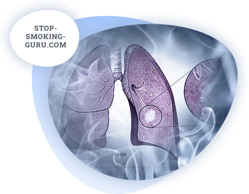
Cancer develops somewhat more frequently in the right lung (51.4%) and less frequently in the left lung (48.6%). The upper lobes are affected more often (60%).
Macroscopically, according to the type of tumor growth, exophytic (endobronchial), when tumor grows in the lumen of bronchus, and endophytic (exobronchial), when tumor grows in the side of lung parenchyma.
There are also intermediate forms of growth.
Microscopic structure of lung cancer. Cancer develops from metaplasized bronchial epithelium and bronchial glands. Pathologically distinguished:
- Squamous cell keratinizing and non-keratinizing cancers-the most common forms (60%);
- Undifferentiated cancer - large-, small-, oat-cell cancer, developing (30%) more often in young, early and widely metastasized;
- adenocarcinoma (10% of cases), consisting of differentiated, slow-growing cells, and undifferentiated adenocarcinoma, with rapid growth, early and widely metastatic.
The anatomic features of the lung structure and the richly presented lymphatic vessels create favorable anatomic conditions for diverse, abundant, and early metastasis of malignant tumor. Lung cancer metastasis occurs by lymphogenic and hematogenous ways. There are three lymphatic barriers on the way of lymph outflow from the lung: pulmonary lymph nodes, located in the thickness of the lung along the bronchi and vessels; bronchopulmonary and tracheobronchial (right and left tracheobronchial and bifurcation) lymph nodes.
There may be some regionality in lymph outflow from the lobes of the lung, but when the overlying lymph nodes are blocked by the tumor process, the lymph flow often goes retrograde. Therefore, it is often necessary to remove all three lymphatic barriers as regional areas of metastasis during surgeries.
Distant metastases are observed in the cervical, supraclavicular and para-aortic lymph nodes. In later cases - in liver, bones, brain and other organs.
Knowledge of the character of tumor growth, histological structure and possible ways of metastasis is necessary for a doctor to understand the peculiarities of clinical picture of lung cancer, to be able to make a diagnosis.
Classification of lung cancer. Domestic classification
Stage I. Small limited tumor of large bronchus endo- or peribronchial form of growth, and also tumor of small and smallest bronchi without pleura invasion and without signs of metastasis.
Stage II. The same or larger tumor, but without pleural leaflet invasion, with solitary metastases in the nearest regional lymph nodes.
Stage III. Tumor that has grown beyond the lung, grows into one of the adjacent organs, pericardium, mediastinum, chest wall, diaphragm in the presence of multiple metastases in the regional lymph nodes.
Stage IV. Lung tumor with extensive spread to adjacent organs, with extensive metastases, distant metastases.
TNM classification of lung cancer
TO - no primary tumor is detected.
TX - presence of a tumor is proved by the presence of cancer cells in sputum, secretion. On radiological or endoscopic examination, the tumor is not detected.
Tl - a tumor measuring 3 cm or less in large diameter, not infiltrating the lobular bronchus (by bronchoscopy).
T2 - tumor more than 3 cm in the largest diameter or tumor of any size with atelectasis or obstructive pneumonitis spreading to the root zone. On bronchoscopy, the spread of the visible tumor must be at least 2 cm distal to the carina. Associated atelectasis or obstructive pneumonitis involving less than a whole lung without pleural effusion.
TZ - Tumor of any size spreading to adjacent structures (mediastinum, thorax, diaphragm) or tumor on bronchoscopy less than 2 cm distal to the carina, or tumor combined with atelectasis or obstructive pneumonitis of the whole lung or pleural effusion.
N - regional lymph nodes.
N0 - no metastases to regional lymph nodes. N1 - metastases to the lymph nodes of the root of the lung side of the lesion, including direct spread of the primary tumor.
N2 - metastases to mediastinal lymph nodes.
M - distant metastases.
MO - no distant metastases.
Ml - distant metastases, including lymph nodes pre lung, cervical, subclavian, opposite lung root, and metastases to other organs.
Clinic of lung cancer
Clinical picture of lung cancer is rather various and depends on localization, stage of disease, anatomic type of tumor growth, histological structure, and preceding lung cancer diseases.
Clinical and anatomical classification of lung cancer according to A.I. Savitsky:
- Central cancer (endobronchial, peribronchial, branched) is the most common form, developing from the epithelium of the main, lobe, segmental and subsegmental bronchi.
- Peripheral cancer (globular, pneumonia-like, lung apex cancer (Penkosta), develops from the alveolar epithelium or epithelium of small bronchi.
- Atypical forms (depending on the features of metastasis - mediastinal, carcinomatosis of the lung, bone, brain, liver, etc.).
One of the basic and the earliest symptoms of central lung cancer is cough, at first dry, at a later stage - haunting, especially at night, accompanied by sputum with blood admixture in the form of streaks, that is a sign of tumor decay. Then there is chest pain, most often due to pleural infiltration of the tumor or in connection with atelectasis and nonspecific pleurisy.
As the tumor grows, the bronchial lumen is narrowed and ventilation of the lobe or segment of the lung is impaired. At first, hypoventilation develops, followed by atelectasis and associated pneumonitis. The latter is characterized by sudden rise in body temperature, increased cough. The clinical picture may be dominated by such general symptoms as malaise, unmotivated weakness, rapid fatigue, reduced ability to work, weight loss, subfebrile temperature. Sometimes there is a hypertrophic pulmonary osteoarthropathy - Marie-Bamberger syndrome, consisting in hypertrophy, calcification and ossification of the periosteum of small tubular bones, forearms, tibia. Clinically, it is manifested by thickening of the tips of the fingers, taking the form of "drumsticks", pain in the limbs especially in the joints. However, this syndrome is not specific and may occur in cases of various metabolic disorders (often with chronic lung and heart diseases).
In contrast to the central cancer, the clinical picture of peripheral cancer is relatively poor. Developing in peripheral zone of lung, far from large bronchi, not causing any evident reaction of lung tissue, a tumor for a long time (in some cases for several years) has no clinical symptoms and is found accidentally during X-ray examination. The first clinical symptom is chest pain on the side of the tumor which appears as pleural effusion, dyspnea and its degree of intensity depend on the size and position of the tumor.
Coughing and hemoptysis occur if the tumor invades the bronchi.
Peripheral cancer is more often accompanied by dissemination of the tumor through the pleura and formation of pleural exudate. The pneumonia-like form of peripheral lung cancer has symptoms much like pneumonia.
Apex lung cancer (Penkosta tumor) due to the predominant spread of tumor infiltrate through the pleural dome to the posterior segments of 1 - 11 ribs, sometimes the arches of the lower cervical vertebrae and to the sympathetic nerve trunk results in a unique clinical symptomatology, in which the pain in the shoulder joint, shoulder and Gorner syndrome prevail.
The clinical picture of atypical forms of lung cancer is caused by metastases, and the primary focus in the lung cannot be identified by the available diagnostic methods. Mediastinal form of lung cancer is distinguished due to peculiarities, caused by predominant growth of metastatic tumor nodes in mediastinum. It is characterized by a pronounced compression syndrome, the degree of which depends on the localization and size of the corresponding groups of lymph nodes. When the lymph nodes around the superior vena cava, anamniotic and neparietal veins are affected, compression symptoms prevail (puffiness of the face, neck, dyspnea, cyanosis, dilation of the subcutaneous venous network on the chest wall).
Predominantly left-sided location of tumor nodules in the mediastinum causes compression paresis of the recurrent branch of the vagus nerve with a hoarseness of the voice up to aphonia. When the posterior mediastinal lymph nodes are affected, symptoms of sympathetic nerve compression (pain syndrome) and esophageal compression (dysphagia) are possible.
Atypical primary lung carcinosis is characterized by multiple focal lung metastases, against which the primary focus is usually not detected. Clinically, it is manifested by dyspnea due to the lesion of a large respiratory surface of the lungs.
Symptoms of lung cancer, connected with tumor invasion of anatomic formations, as well as regional and distant metastasis, are very diverse.
Lung cancer diagnosis is difficult at all stages of tumor development. In the early period there are no symptoms, in the later stages there are great difficulties in differential diagnosis. Detection of the early stages of globular peripheral lung cancer is possible with prophylactic fluorography. In other cases, the possibility of early diagnosis is associated with some form of dispensary observation. This work requires the participation of highly qualified specialists, and detailed specifying examination of patients can be carried out in highly specialized broncho-thoracic diagnostic departments. First of all, pulmonology advisory (oncology) commissions are organized from competent specialists (statisticians, oncoepidemiologists, cytologists, pulmonologists, surgeons, radiologists, etc.) to form oncorrisk groups.
The latter are organized using the questionnaire method. Risk factors for lung cancer should be considered: age over 45 years for men and 50 years for women, occupational hazards (gas and petrochemical industry, miners, asphalt workers, cement, asbestos, chrome, etc.), tobacco smoking, chronic inflammatory lung diseases, etc. People of high risk group should be examined at least twice a year by a sputum total analysis with microscopic examination for definition of the cellular structure with signs of atypia (cytological screening). As practical experience of leading medical institutions of our country shows, in 3 - 15% of those registered in the risk group the IV degree of cell atypia is diagnosed. These individuals require additional clarifying examination in specialized diagnostic broncho-thoracic departments.
In later stages, primary diagnosis is made on the basis of history taking, physical, laboratory, radiological and endoscopic examinations. Particular attention should be paid to patients with prolonged cough, hemoptysis, and recurrent fever. On physical examination, the presence of restriction of chest mobility, areas of shortened pulmonary sound, and weakened breathing should be noted. In all cases, multiple sputum examinations for atypical and tumor cells are important to confirm the diagnosis.
The main method of recognition of lung cancer is radiological. In peripheral lung cancer, radiographs show a round, dense shadow of a tumor with rather clear, bumpy contours. In the presence of concomitant inflammatory processes in the lungs, diagnosis is difficult. Central cancer is characterized by symptoms of hypoventilation or atelectasis of the whole lung, lobe or segment. However, most often it is a manifestation of a far advanced process that is not subject to radical surgical intervention.
Diagnostics of earlier forms of the disease turns out to be difficult. In these cases of clinical and radiological symptomatology of lung cancer with specific and nonspecific inflammatory processes a special oncological alertness of a general practitioner is required. Relying on fluoroscopy and single projection radiography data, being under the impression of radiological diagnosis, the clinician does not always correctly make clinical and radiological parallels, does not insist on more complete pulmonological examination of a patient, including a thorough tomographic examination of the lung, bronchial tree and mediastinum.
Tomograms allow to obtain clearer outlines of a primary focus in peripheral lung cancer, especially in case of a pronounced paracarcinoma inflammatory reaction "covering" the primary tumor. At central lung cancer, tomography reveals indistinctness, deformation and usurpation of bronchial walls, narrowing of bronchial lumen, presence of enlarged lymph nodes in the area of roots and mediastinum. Computed tomography is an extremely informative method of examination.
Examination ends with endoscopic examination with biopsy of the tumor or, in case of peripheral cancer, catheterization of small bronchi, with taking material for cytological examination. If necessary, perebronchial puncture of primary focus and enlarged lymph nodes or transthoracic puncture of subpleural tumor is performed in a special diagnostic broncho-thoracic department. In some patients final diagnosis can be established only by diagnostic thoracotomy.
X-ray examination under superimposed pneumothorax, pneumomediastinotomography, bronchography are used for specifying diagnosis and revealing the extent of process prevalence.
Additional data can be obtained using angiography, including azigography. Parasternal mediastinotomy, biopsy of enlarged peripheral nodes, mediastinoscopy with biopsy, fibrobronchoscopy with peritracheal and perebronchial lymph node puncture, cytological examination of pleural fluid, laparoscopy and laparotomy are also used.
The introduction of special methods of determining the degree of spread of the tumor process promotes operability and resectability, decreases the number of trial thoracotomies, increases the number of radical operations, improves planning of rational conservative therapy and improves treatment results.
Principles of lung cancer treatment
At present time at treatment of lung cancer we use surgical, radiological and chemotherapeutic methods of treatment or their combinations in various combinations. The choice of treatment method depends on the localization, clinical and anatomical forms of the tumor, the stage of its histological structure and the degree of cell differentiation, functional abilities of a patient, especially respiratory function and cardiovascular system. Surgical treatment turns out to be effective in stages I, II and sometimes III in squamous cell lung cancer and adenocarcinoma. At undifferentiated lung cancer preference should be given to radiotherapy and drug treatment. Among operations there are typical (lobectomy, bilobectomy, pneumonectomy) and combined operations, which are accompanied by resection of the adjacent anatomical formation (part of the diaphragm, chest wall, pericardium).
If localization of central lung cancer allows to cut off the lobar bronchus, with at least 2 cm from the tumor edge, in the absence of metastases in the root and tracheobronchial lymph nodes, lobectomy is indicated. There are greater opportunities for this in peripheral cancer.
In all other cases pneumonectomy with removal of bifurcation, tracheobronchial lymph nodes with fatty tissue on the side of the lesion is indicated.
In low-differentiated forms of cancer, pneumonectomy is indicated because of frequent and more extensive metastasis to regional lymph nodes.
For more details on surgical treatment of lung cancer see: B. E. Peterson, Surgical Treatment of Malignant Tumors. Medicine, 1976, 222 - 237).
In recent years the combined method including preoperative radiation therapy and radical surgery is more promising.
Preoperative radiation therapy is aimed at reducing the biological activity of the tumor, reducing its size, destroying subclinical metastases in regional lymph nodes. As a result, a decrease in the rate of recurrence and metastasis is achieved. The irradiation is usually performed using modern gamma and light particle gas pedals with 15-25 MeV energy in medium fractions of 5 Gy daily for 5 days until the total focal dose of 25 Gy, and the surgical intervention is performed 2-4 days after the end of radiotherapy. In cases where there are doubts about the radicality of surgery, postoperative radiation therapy is resorted to; it is usually begun 3 to 4 weeks after surgery with a total focal dose of 30 to 40 Gy and a daily dose of 2 Gy.
Radiation therapy as an independent method can be performed according to a radical or palliative program. The main indication for radiation therapy on the radical program is a relatively small tumor, with a satisfactory condition of patients (stage 1 - 2 of the disease), who refuse surgery, or not subject to surgical intervention due to general contraindications. Usually the irradiation is performed through shaped fields with capturing the primary tumor, as well as subcorneal, paratracheal and bifurcation lymph nodes in the irradiated zone. Areas of atelectatically changed lung are not included in the irradiation area. In undifferentiated forms of cancer the supraclavicular areas, the whole mediastinum, the area of the opposite root are included in the irradiation zone. Weekly dose is 10 Gy. Total focal dose is 60-70 Gy.
Palliative radiotherapy is carried out on patients with stages III and IV, when there is metastatic lesion of all groups of regional lymph nodes or spread of tumor to ribs and pleura, with the involvement of great vessels, with single metastases in supraclavicular region. The total focal dose is 45-50 Gy.
The technique of radiotherapy by a split course was widely used. After a total focal dose of 30 Gy has been administered, a break is made for 3 weeks.
After a total focal dose of 30 Gy is applied, a break is made for 3 weeks, after which another 40 Gy is applied to the focus. This method has better tolerability of palliative treatment without reducing the effectiveness of radiotherapy. Contraindications to radiation therapy are advanced age, cachexia, concomitant active pulmonary tuberculosis, tumor decay, bleeding, distant metastases, etc.
You can read more detailed information about radial therapy and combined treatment of lung cancer in the book "Radial therapy of malignant tumors" by A.V. Kozlov, Moscow, "Medicine", 1976, pp. 112 - 118. 112 - 118.
Recently in clinic and outpatient treatment of lung cancer antitumor chemotherapeutic agents have been more and more widely used.
Opportunities of chemotherapy of lung cancer are determined first of all by histological type of a tumor. In this connection, small cell anaplastic lung cancer of many smokers of 40 - 45 years old is distinguished by its extremely malignant course, propensity for early generalization of process, and at the same time high sensitivity to chemotherapeutic (and radiation) influences. Other histological types - epidermoid squamous cell, large cell and adenocarcinoma are less sensitive to chemotherapeutic agents.
At treatment of small cell lung cancer the following schemes of polychemotherapy (combined chemotherapy) are applied: cyclophosphane, adriamycin, methotrexate; cyclophosphane, adriamycin, vincristine; adriamycin, cyclophosphane, etoposide etc. Recently, there is evidence that the therapeutic effect, reduces the number of local relapses.
Polychemotherapy for non-small cell cancer has little effect.
Somewhat greater sensitivity is observed in low-differentiated forms of cancer.
Use of cytostatic agents is of some benefit in exudative pleurisy complicating the course of lung cancer. Repeated intrapleural administration of thiophosphamide in single doses of 30-40 mg after maximal evacuation of pleural cavity contents in 60-70% of cases result in pleural cavity obliteration and termination of exudate accumulation in patients with various histological forms of lung cancer.
Intrapleural administration of cyclophosphan or bleomycin gives similar effect.
For a more detailed introduction to the topic of chemotherapy for lung cancer, see:
- Blokhin N. N. N., N. I. Transluchikova, "Chemotherapy of Tumor Diseases," Moscow, Medicine, 1984, 163 - 180.
- Transluchikova N. I. "Antitumor chemotherapy," Moscow, "Medicine," 1986, 44 - 48.
Treatment outcomes and prognostic factors
With surgical treatment, the 5-year survival rate can be achieved in only 25-40% of operated patients. Unsatisfactory results of surgical treatment are most often related to the nonradical nature of the operation. Longevity of a patient is influenced by the extent of spread of process, morphological structure of lung tumor and differentiation degree.
Thus, at the I stage 48.5% of patients live more than 5 years, at the II stage - 41.3%, and at the III stage - 18.4%. In the presence of squamous cell lung cancer 34% of patients live more than 5 years, and glandular - 33.3%. In undifferentiated cancer the survival rate was only 7.7%.
Radiation therapy can improve the condition of patients, prolong their life. Average life expectancy of patients after radiation therapy is 16.7 - 18.9 months, reaching 2 - 3 years, and sometimes 5 years (10%).
Combined chemotherapy in small cell lung cancer allows to achieve a significant direct effect in 75% of patients, and in 25-45% of patients complete disappearance of all clinical signs of the disease is achieved. The average life expectancy is 8 - 13 months.
Only patients who manage to achieve complete clinical remission have real chances for prolongation of life up to 2 years and more.
Improvement of results of treatment of this severe disease is connected with improvement of modern diagnostics, by active detection of lung cancer among the population "at risk",
Radiographic diagnosis of lung cancer
Lung cancer (bronchogenic carcinoma) has become one of the most formidable human diseases in recent years. In its frequency in industrialized countries it is second only to malignant illnesses of the stomach. The share of lung cancer among oncological diseases in Russia is about 15.0%. At the same time it is the most widespread localization of cancer in men. In 35 industrial countries of the world the bronchogenic carcinoma is the main cause of death among oncological patients.
The annual increase in the incidence of lung cancer for the whole population of the Russian Federation reaches 3.5%. The spread of this form of cancer has become epidemic. In the United States medical statistics reveals a no less dramatic picture. So, in 1912 there were 394 new cases of bronchogenic carcinoma, in 1981 - 140,000, in 1991 - 161000 and in 1993 - 169900 cases. Given the steady increase in incidence, another 200,000 new cases of lung cancer are expected to be found in the United States in 2000. The established fact can no longer be explained only by improved diagnosis and a demographically aging population.
Lung cancer is most often detected in the seventh decade of life, with men suffering five times more often.
According to clinical and anatomical classification of lung cancer accepted in Russia the following forms of cancer are distinguished.
Central: a) endobronchial; b) peribronchial nodular; c) branched.
Peripheral: a) circular shadow; b) pneumonia-like; c) apex of the lung (Pancost).
Atypical forms: a) mediastinal; b) miliary carcinomatosis.
International histological classification of lung tumors was published in 1981. They distinguish squamous cell cancer, small cell cancer, adenocarcinoma, large cell cancer, glandular squamous cell cancer, carcinoid tumor, bronchial gland cancer and other forms.
Squamous cell cancer is the most common - according to various studies up to 50 - 75%, and with age, its proportion increases to 78%. Men get squamous cell cancer almost nine times more often than women.
Like other types of tumors, lung cancer is increasingly being detected at a young age. This is primarily due to an increase in the proportion of adenocarcinoma.
Among the main radiological methods used to detect lung cancer are fluoroscopy, chest radiography, computed tomography, and, less frequently, magnetic resonance imaging. Additionally, fluoroscopy, bronchography, angiography, and diagnostic pneumothorax may be used.
Chest fluorography has become widespread in Russia. This technique was optimal in the period of widespread introduction of mass clinical examination of the country's population. However, it is necessary to admit its main drawback: the low level of sensitivity and accuracy. Experience shows that the rate of false positive and false negative findings reaches 30%. Besides, the radiation exposure of patients and medical personnel is unreasonably high. Absorbed dose during fluorography is more than 0.25 mSv.
Chest radiography has better resolution and lower radiation exposure (0.05 mSv). Clinics in Western Europe and North America are aggressively adopting digital technology and filmless production. Digital radiography further reduces the radiation load on the patient. However, the appropriateness of its use in pulmonology is still debatable. The arguments of supporters of analog images have a strong point - the resolution of the latter in the study of the lung pattern is higher.
Digital technologies are constantly developing. The emergence of flat panel detectors promises to revolutionize radiography. On the one hand, their resolution is higher than the usual analog images, and on the other hand, the radiation load is expected to be reduced by up to 75%. Therefore, digital radiography should become a screening examination of chest organs in the coming decades.
Lung cancer in 60% of patients has central localization. It is known to develop from the epithelium of the segmental bronchi. Then the tumor grows proximal to the main bronchus. More often, segments III and VI of the right lung are affected.
Semiotic signs of central lung cancer on radiography include:
- The presence of tumor masses in the root region of the lung;
- hypoventilation of one or more segments of the lung;
- signs of valve emphysema of one or more lung segments;
- atelectasis of one or more lung segments.
X-ray diagnosis of peripheral lung cancer is often difficult due to the similarity of its semiotic signs with other diseases. The problem is not in the detection of the mass, but in the interpretation of the obtained data. A single lesion of the upper lobe of the lung is typical. The structure is more often homogeneous, but in fact necrosis occurs quite often. However, bronchial drainage remains unrecognized for a long time. The timing of tumor doubling depends on the histologic structure of the tumor. Thus, in squamous cell cancer, it averages 140 days.
Apex cancer is accompanied by Pancost syndrome. It is characterized by the presence of a rounded mass in the area of the lung apex, pleural changes, destruction of the upper ribs and corresponding vertebrae. Pancost's cancer can only be talked about when it has a true bronchogenic origin.
An uncommon complication of peripheral lung cancer is decay of the tumor node. At pseudocavernous form of cancer along with already mentioned semiotic signs there are additional features helping to distinguish it from abscess. Decay cavity can occupy a considerable part of tumor masses. At the same time the cavity has uneven, festooned character. Clinically, there are no signs of lung abscess.
Unfortunately, as in most cases, radiography does not allow to determine the histological nature of the disease. However, there are some patterns to consider when analyzing the images. Thus, adenocarcinoma more often has peripheral localization in the upper lobes of the lungs and is solitary. The structure, as a rule, is heterogeneous, lobular. Contours are uneven, festooned.
Squamous cell carcinoma is primarily characterized by local thickening of the bronchial walls, subcortical location and, therefore, by the occurrence of atelectasis. Squamous cell carcinoma is prone to internal decay.
Squamous cell carcinoma is also more often central. The primary tumor is detected radiographically very rarely. At the same time, mediastinal lymphoadenopathy is characteristic.
Large cell carcinoma may have any localization. At the same time, in 70% of patients tumor masses at primary detection already exceed 4 cm in diameter.
Carcinoid tumor is uncommon and primary malignant. In 80% of patients it grows endobronchially and solitarily at lobular, segmental, or subsegmental level. In one-third of cases, abnormalities of the bronchial tree are additionally visualized.
Bronchial gland cancer has a central localization in 85-90% of cases. In this case, it originates from the trachea or main bronchi and leads to their narrowing.
The second mandatory step in the examination of patients with suspected lung cancer should be an X-ray computed tomography (CT) scan. Today computed tomography has firmly and indisputably taken its place among routine examinations of such patients in many Russian clinics. This is due to the high sensitivity of the technique. Despite the impossibility of histological verification of the tumor, the specificity of RCT is also higher than that of radiography, reaching 70%.
The standard protocol of computed tomography-examination includes consecutive scans with a slice thickness and a table feed step of 10 mm. If necessary, the spatial resolution can be improved by using 2-mm-thick "slices" in a specific area of interest. For example, this is required to confirm the presence of calcinates. Scans of 5 mm thickness are optimal in the study of lung roots to determine lymphadenopathy and study of a central lung cancer node.
The study can be performed in both stepped and spiral RCT modes. The advantages of the second technique in lung investigation are:
- possibility of examining the whole zone of interest with one or two breath-holds;
- Exclusion of factor of different respiratory depth during examination, which increases sensitivity of the method.
Intravenous contrasting with iodine-containing agents (Omnipack, Ultravist) is not often used in step tomography. The accumulation of contrast agent in the tumor tissue usually does not provide additional diagnostically significant information.
This is not the case with spiral CT. A fundamentally new possibility of bolus contrasting allows the study to be performed at 25-30 seconds of introduction of the drug. In this case 50-60 ml of drug is injected into the cubital vein at a rate of 2 ml/s. Identification of vascular structures and their topographic and anatomical relationship with neoplastic masses significantly improves. Thus, involvement of vessels of lung root and mediastinum is determined.
Analysis of tomograms is performed in two modes: mediastinal and pulmonary. In the first one, soft tissue structures are studied in detail with window width from 0 to 1000 HU and window center 50 HU. In the second one, the subject of investigation is air pulmonary tissue with window width from 1500 to 500 HU and window center - 650 HU. Normal densitometric values of lung tissue are about 760 HU. At the same time they are slightly higher in the lower sections (by 80 - 100 HU), which is explained by gravitational influence on blood placement in the vascular channel.
The most radical way to treat lung cancer remains surgery. Computed tomography allows to specify the risk and justification of surgical intervention, to estimate the possibility of complications, to determine the prognosis of patient's treatment.
When examining a patient with lung cancer X-ray computed tomography has the following objectives:
- to clarify the presence and solitariness of the formation, to identify possible metastases;
- study the size, structure, density (quantitatively in HU) and contours of the tumor;
- to define relations of neoplastic masses with surrounding morphological structures of lung root and mediastinum;
- to specify possible origin of the tumor;
- to study changes of lung tissue;
- reveal involvement of mediastinal lymph nodes.
Typically, central cancer is characterized by a polymorphic structure, a homogeneous structure, and a density of 40 to 70 HU. Foci of decay tend to appear when the tumor is of significant size.
Computed tomography allows fairly accurate identification of the root of the lung and the root mass.
Central cancer of the upper lobe bronchus of the left lung, exogenous growth:
- Tracheal bifurcation level. Mediastinal view. A tumor spreading into the mediastinum, irregular round shape, heterogeneous structure, density 40 - 55 HU, with lumpy contours is visualized. A group of lymph nodes up to 1.5 cm in size was detected against the mediastinal fatty tissue.
- The level of the pulmonary artery trunk. Double window mode.
Tumor masses concentrically surround the upper lobe bronchus of the left lung, intimately adjoining the left pulmonary artery
Often, pneumonia on radiographs can conceal central bronchial obstruction. RCT is helpful in detecting it. Problems arise only when the densitometric indices of tumor and inflammatory lung infiltration are close in value.
Detailed analysis of consecutive scans helps in determining the localization of bronchial obstruction.
In terms of resectability, the relationship of tumor with surrounding morphological structures is important. There is a high probability of involvement of mediastinal organs, vessels, pleura, chest wall in neoplastic process. A good natural contrast background is fatty tissue with density from 90 to 80 HU.
There are significant advantages of computed tomography over traditional radiography in assessing the degree of obstructive syndrome. In particular, the stage of hypoventilation can be detected earlier than in radiography.
Central cancer of the lower lobe bronchus of the right lung, exogenous growth. Hypoventilation of the ninth segment of the right lung:
- The level of the pulmonary artery trunk. Infiltration of mediastinal fatty tissue. Minor right-sided pleurisy.
- ThIX vertebral level. Double window mode. The right lower lobe bronchus is pushed backward by tumor masses. Decreased pneumatization, enrichment of the lung pattern in segment IX of the right lung.
It is characterized not only by enrichment of lung pattern and signs of volumetric reduction, but also by increase of density (decrease of pneumatization) of affected lung segment (or lobe) up to 400 - 600 HU. On radiographs, changes are visualized only with increase of lung tissue density up to 250 - 200 HU.
Computed tomography is less informative in atelectasis development. Only in 20-25% of patients it is possible to clearly differentiate the tumor borders on the background of dense atelectasis. Densitometric indices of atelectasis sites vary from -10 to 40 HU.
Peripheral squamous cell cancer of segment VI of the right lung. Pulmonary mode. A rounded tumor, up to 1.8 cm in size, of homogeneous structure is visualized. Signs of regional lymphangiitis in the form of "radial crown" and "track" to the lung root and pleura. Additionally, subpleural metastasis of the right lung is detected paravertebral
Always during chest CT of the organs, lymph nodes of the mediastinum and lung roots are evaluated. Many authors are skeptical about the role of CT in detecting lymphadenopathy. Indeed, if we proceed from the idea that lymph node sizes can normally reach 1.0-1.5 cm, the specificity of the technique is low: about 40% in central cancer and 60-65% in peripheral cancer. Excellent visualization of lymph nodes up to 3 mm in CT does not allow to state their possible affection. At the same time specificity of investigation, according to the majority of scientists, can reach 90-97% if only nodes increased up to 1.0 cm and more are considered.
A complication of central lung cancer can be effusion pleurisy, the severity of which depends on the degree of pleural involvement. Computed tomography reveals a minimal, 10-15 ml, amount of fluid in the pleural cavity.
X-ray computed tomography is certainly more sensitive and specific for detecting peripheral tumors.
Peripheral squamous cell cancer of segment VI of the right lung with pleural involvement. Bilateral pleurisy:
- Patient position on the back. The "double window" mode.
- Patient position on the abdomen. Pulmonary mode. Fluid moved freely. Better visualized signs of regional lymphangiitis.
However, more often its role is reduced to the search for similar masses in other parts of the lungs and other organs. That is, the solitary nature of the neoplastic process is confirmed.
Peripheral cancer of the lower lobe of the right lung, infiltrative form, with involvement of the pleura. Moderate right-sided pleurisy
The most reliable sign of malignancy of a mass is an increase in the size of the node. Homogeneity of the structure also indicates in favor of cancer, although small calcinates and foci of decay may occur. The third sign - regional lymphangiitis - significantly objectifies the diagnosis of tumor. It manifests as "radial crown" and "track" to lung root and pleura. As it progresses, there is perifocal reduction of pneumatization of parenchyma. With involvement of visceral pleura leaflet in pleural cavity a small size effusion is detected, which is not revealed even by laterography.
X-ray computed tomography is more sensitive in detecting the infiltrative form of cancer, apical cancer, and tumor decay.
Apical cancer (adenocarcinoma) of the right lung. Mediastinal mode, consecutive scans. Infiltration of pulmonary tissue and mediastinal fatty tissue is defined around the tumor masses.
Peripheral cancer of the lower lobe of the left lung, with decay and involvement of the pleura. Double window mode.
Almost all round tumors cause diagnostic difficulties to some extent. Peripheral lung cancer has to be differentiated with a number of diseases:
Neoplasms:
- malignant;
- Bronchoalveolar cancer, sarcoma, metastases;
- benign.
Inflammatory lung diseases:
- Cysts.
- Pleurisy.
- Pleural tumors.
Primary involvement of ribs, vertebrae.
Primary involvement of diaphragm.
Bronchoalveolar cancer of the right lung, diffuse form.
Multiple hematogenous metastases of both lungs.
At the same time, in the majority of cases only histological examination makes it possible to convincingly testify in favor of this or that lung disease. Therefore, fine-needle biopsy is widespread in the world. Unlike other internal organs, biopsy of lung masses is possible only under the control of RCT. The disadvantage of such control method, in contrast to ultrasound scanning, is time delayed image acquisition.
Thus, the problem of early diagnostics of lung cancer is far from being finally solved. However the development of modern high technologies, their implementation in medicine open up new possibilities of detecting tumor process.
Gamartoma of the VI segment of the right lung.
Gamartochondroma of the right lung.
Mesothelioma of the right lung. Mediastinal mode:
- Right pulmonary artery level.
- Level of the base of the heart.
Pleural thickening is detected, with the main tumor nodule at the level of the base of the heart. Expressed right-sided pleurisy.
Ubiquitous introduction of digital roentgenography and computed tomography in Russia will allow to increase the proportion of lung tumors diagnosed at the initial stage of the process already at the beginning of the new century. Low specificity of radiological methods of examination will be compensated by the use of fine-needle biopsy of neoplasms under control of RCT.
Thin-needle biopsy of a peripheral volumetric mass of the upper lobe of the left lung under the control of CT. Puncture was performed through the anterior chest wall. The end of the needle was defined in the center of the tumor.
In recent years technically the task of computed tomography in fluoroscopic mode has been solved. The latter will allow to perform invasive manipulations (blockages, punctures, biopsies) under the control of RCT in real time. So far, only a few RCT machines in Russia have the necessary software for fluoroscopy. This is another promising area of radiodiagnostics development that is expected to grow in the next few years.
Pay attention! X-ray diagnosis of peripheral lung cancer is often complicated due to the similarity of its semiotic features with other diseases.
The obligatory stage of examination of patients with suspected lung cancer should be X-ray computed tomography.
The specificity of computed tomography is higher than that of radiography and reaches 70%.
Only histological examination is still the most accurate diagnostic method. Currently, fine-needle biopsy under the control of computed tomography is widespread.
Computed tomography allows to specify the risk and determine whether surgical intervention is justified, assess the possibility of complications, and build a prognosis for the patient's treatment.


Lung Cancer
Etiology and statistics
Lung cancer is one of the most common malignancies. The predisposing factors have been studied in great detail. The main role in the etiology of the disease most researchers attribute to long-term inhalation of air polluted with various carcinogenic substances. The most potent of these substances is 3,4-benzpyrene. Of importance is the ingestion of carcinogenic substances into the lungs with adsorbents, such as dispersed soot, which contributes to their fixation in the tissues. Inflammatory changes, atrophic bronchitis, various dust diseases, in which the mechanism of physiological self-purification of the bronchopulmonary system (L.M. Shabad) is disturbed, can also contribute to the same. The undoubted influence of smoking on the incidence of lung cancer has been established. According to data published in the USA, people who smoke more than two packs of cigarettes a day have 20 times higher risk of getting lung cancer than nonsmokers. There is evidence that cigarette smoking is more closely linked to lung cancer than cigar or pipe smoking. Cigarette tobacco has a higher carcinogenic arsenic content, and cigarette smokers tend to inhale more smoke into their lungs.
Special studies in various countries have found an undeniable and continuing increase in the incidence of lung cancer. This increase is true, i.e., not related to increased longevity of people and improved diagnosis. In the USA in the same age groups of the population from 1914 till 1950 the death rate of men from lung cancer has increased in 28 times, and in women - in 7 times. Among the urban population of Ukraine, mortality from lung cancer in the same age groups increased 3-fold among men and 2-fold among women.
To explain this increase in the incidence of lung cancer, various concepts have been proposed. The main attention is paid to two factors, the great importance of which is generally recognized.
The first factor is an increase in atmospheric air pollution, which in all industrialized countries has increased in recent decades due to increased emissions of products of incomplete combustion of coal, fuel oils, gasoline, various gases and dust, such as that produced when car tires rub against bituminous road surfaces.
The second important factor is the increase in tobacco consumption. In urban populations and especially in large, smoky cities, lung cancer is more common than in rural areas. Men, especially those over 40 years of age, are much more likely to get lung cancer than women. More than half of lung cancer patients are heavy smokers.
Pathologic anatomy
Lung cancer usually develops from the epithelium of the bronchi and much less often from the epithelium of the alveoli, so it is usually a bronchogenic cancer. In most cases, the cancer originates in the segmental and subsegmental bronchi, and then, as it grows, it invades the larger lobular and major bronchi. The right lung is affected slightly more often than the left lung, and the upper lobes of the lungs are affected more often than the lower lobes. In the upper lobes it develops more frequently in the anterior segments, and in the lower lobes in the apical segments.
Cancerous tumors arising from large bronchi of segmental, lobe, and major bronchi are usually closer to the root of the lung and are therefore called central, and those arising from smaller ones are called peripheral. This division is of great practical importance, because the clinical manifestations and diagnostic principles of central and peripheral cancers are different.
In case of central cancer it is advisable to distinguish the character of tumor growth: mainly in the side of bronchial lumen (endobronchial) or outside it - in the side of lung (peribronchial). The form of growth in both cases can be either nodular or branching. Peripheral cancer usually presents as a tumor nodule of globular or spherical shape. Sometimes the growth process of peripheral lung cancer proceeds in parallel with the destruction of lung tissue and the formation of a cavity. Such cancer is called primary cavity cancer. There are also known cases of infiltrating tumor growth without definite form and clear borders (pneumonia-like cancer).
According to histological structure, a distinction is made between differentiated, which occurs more often, and relatively rare undifferentiated lung cancer. Epidermoid cancer and carcinosarcoma belong to differentiated cancer, small cell cancer and polymorphous cell cancer belong to undifferentiated cancer.
At the initial stage of development, of course, the cancerous tumor is small and barely visible to the eye, but in advanced cases it can occupy almost the entire lung, grow together with multiple metastatic nodes and infiltrate surrounding organs and tissues. Lung cancer metastasis occurs lymphogenically and hematogenously.
The first stage of lymphogenic metastasis is pulmonary and bronchopulmonary lymph nodes located along segmental and lobe bronchi. Tracheo-bronchial, bifurcation, paratracheal and mediastinal lymph nodes are further affected. When regional lymph nodes are blocked, retrograde outflow of lymph occurs, resulting in possible tumor metastasis outside the normal lymph outflow pathways. Hematogenous metastasis of lung cancer occurs to the liver, brain, bones (spine, ribs, skull), adrenal glands, kidneys, rarely to the pancreas, spleen, opposite lung, thyroid gland, skin. Such distant metastases occur more frequently in younger patients and especially in cases of undifferentiated forms of cancer, which metastasize earlier and more intensely than differentiated tumors.
Classification
The size and prevalence of the tumor, together with the presence or absence of metastases in regional lymph nodes, underlies many classifications of cancer. In the Soviet Union, according to the classification of malignant tumors adopted in 1956, division of lung cancer into 4 stages is widespread: Stage I - small tumor without metastases; Stage II - small or slightly larger tumor with single metastases in bronchopulmonary lymph nodes; Stage III - tumor that has gone beyond the lung, or tumor with multiple metastases in regional lymph nodes; Stage IV - tumor with spread to neighboring organs and extensive regional or distant metastasis. The given division of lung cancer into stages helps to differentiate patients according to the severity of tumor process, but it is seriously behind the modern clinics requirements. Unfortunately, the classification of the lung cancer according to TNM system (T - tumor, N - lymph nodes, M - metastases) suggested in 1965 by the International committee is not satisfactory and that is why it is not widely spread.
In clinical practice the classification of lung cancer based on the suggestions of A.I. Abrikosov, A.I. Savitsky, F.G. Uglov, V.I. Struchkov and reflecting localization, form, character of tumor growth, its histological structure, stage and complications is rational.
Classification of lung cancer
1. Anatomical forms
Central cancer (cancer of the main, lobe and segmental bronchi): 1) with predominantly endobronchial growth; 2) with predominantly peribronchial growth: a) nodular form, b) branching form.
Peripheral cancer:
- nodular,
- cavitary,
- pneumonia-like.
2. Histological forms Differentiated cancer:
- Epidermoid: a) without keratinization, b) with keratinization;
- adenocarcinoma;
- solid cancer;
- carcinosarcoma.
3. Undifferentiated cancer:
- small-cell,
- polymorphic cell cancer.
According to this classification, the detailed diagnosis of the disease looks as follows: epidermoid without keratinization cancer of the right upper lobe bronchus with predominantly endobronchial growth, stage II, atelectasis of the upper lobe.
Symptomatology, clinical course, and principles of early diagnostics are different in central and peripheral lung cancer, especially in early stages.
Central lung cancer
In central cancer, relatively early symptoms may include coughing and slight hemoptysis. Often patients, mostly older smokers, don't pay much attention to these symptoms. As the tumor increases, especially in cases of predominantly endobronchial growth, the affected bronchus narrows - bronchostenosis occurs, which results in hypoventilation of lung segment, lobe and sometimes the whole lung. Quite often during this period a tumor acts as a valve, which doesn't prevent inhalation, but hinders exhalation. As a result, local emphysema appears, which, however, at narrowing of segmental and lobe bronchi usually does not cause subjective sensations and is not determined by methods of physical examination - percussion and auscultation. Only in case of valvular narrowing of the main bronchus and obstructive emphysema of the whole lung patients start to feel dyspnea.
It is possible to suspect central lung cancer during this period only when fluorographic or X-ray examination reveals signs of bronchostenosis in the form of hypoventilation or local emphysema of lung segments or lobes. The presence of these signs is the reason for taking direct and lateral lung X-rays and layer-by-layer tomograms. On tomograms, endobronchial tumors of the main and lobe bronchi are well defined, the contours of tumors and shadows of segmental atelectasis are sharply outlined. The most important and usually decisive methods of investigation for exclusion or confirmation of lung cancer diagnosis are sputum cytological examination and tracheobronchoscopy.
Lung cancer diagnosis
Morphological data (histological and cytological) which confirm the diagnosis of lung cancer and, as a rule, allow determining its histological form, have the major meaning in the diagnosis of lung cancer. Only after their reception the diagnosis of lung cancer becomes verified, i.e. not presumptive or probable, but reliable. In clinical practice, cytological examination of sputum, flushes, scrapes, impressions from the bronchial wall, and aspiration biopsy material during bronchial catheterization is particularly important for verification of the diagnosis. In the presence of radiological changes in the lungs, cytological findings are positive in 85-90% of cases. Sometimes they can be positive even before the appearance of radiological changes in the lungs.
The basic scheme of application of the main methods of diagnostics of the central and peripheral lung cancer seems to be as follows:
- Suspicion of lung cancer
- Radiography and tomography
- Sputum cytology
- Bronchoscopy for morphological examination (for the central form)
- Bronchial catheterization or chest wall puncture with morphological examination (peripheral form)
- Differential diagnosis
In the differential diagnosis of central lung cancer we have to keep in mind chronic pneumonia, tuberculosis, segmental pneumosclerosis, sarcoidosis, lymphogranulematosis, bronchial adenoma. Peripheral cancer mainly needs to be differentiated with benign tumors and cysts of the lung, tuberculomas of the lung, tumors and cysts of the mediastinum, pleura, chest wall and diaphragm, metastases of malignant tumors of other organs into the lungs.
In cases when the diagnosis of lung cancer is very probable, but cannot be verified by various research methods, diagnostic thoracotomy is advisable. With the improvement of lung cancer diagnostics its indications are gradually becoming rarer.
Trachtenberg proposed risk criteria for lung cancer:
1. Genetic factors:
- Primary multiple tumors (previously treated for malignant tumors);
- three or more cases of lung cancer in close relatives.
2. Modifying risk factors:
- Exogenous: smoking, environmental pollution, occupational hazards;
- endogenous: age over 45 years, chronic lung diseases (pneumonia, tuberculosis, bronchitis, localized pneumofibrosis, etc.).
Thus, the decisive role in etiology of lung cancer is assigned to action of carcinogens, and in pathogenesis - to disturbance and perversion of bronchial epithelium regeneration.
Classification. In most cases lung cancer proceeds from bronchi, and the tumor can be located in the central or peripheral zone of the lung. The clinical and anatomical classification of lung cancer, proposed by A. I. Savitsky.
1. Central cancer:
- endobronchial,
- peribronchial nodular cancer,
- branched.
2. Peripheral cancer:
- round tumor,
- pneumonia-like cancer,
- cancer of the apex of the lung (Penkosta).
3. atypical forms associated with peculiarities of metastasis:
- mediastinal,
- miliary carcinomatosis,
- bone,
- cerebral, etc.
Depending on which elements of bronchial epithelium form the tumor, histomorphological forms are distinguished: squamous cell cancer (epidermal), small cell cancer (undifferentiated), adenocarcinoma (glandular), large-cell, mixed, etc.
Lung cancer is a widespread disease. In the structure of morbidity of malignant neoplasms lung cancer in CIS takes the 1st place.
The highest morbidity of lung cancer is registered in New Zealand (Maori), standardized figures make 99,7 0/000; the minimum morbidity of lung cancer in Mali - 5,3 ?/000. In the United States, the absolute number of first-time cases exceeded 100,000 in the late 1980s.
Men fall ill 5-8 times more often than women. The disease is most often registered at the age of 50 and older. Mortality from lung cancer has been decreasing over the last decade (a decrease of 21%).
Contributing factors
There are many causes of lung cancer, they are both exogenous and endogenous. However, exogenous factors are decisive: tobacco smoking, pollution of the surrounding air by carcinogens and radioactive isotopes - both as a result of radioactive contamination and natural natural isotope - radon gas.
Urbanization, growth of industrial enterprises lead to contamination of soil, water, and atmospheric air with carcinogenic substances (3,4-benzpyrene, benzanthracene, etc.).
Smoking tobacco is dangerous because the high temperature of lit cigarettes produces carcinogenic substances (in particular, benzpyrene and benzanthracene), as well as a large number of soot particles (up to 1 million from one cigarette), which act on the ciliated epithelium of the bronchial mucosa, leading to its death. A great role in the mutagenic effect of DNA and RNA, chromosome apparatus of cells is played by radioactive isotope - polonium-210 with a long half-life, which when smoking tobacco is transferred to smoke and lingers in the body for a long time. The synergistic effect of benzpyrene and polonium-210 leads to cancerous cell rearrangement faster. A correlation between the incidence of lung cancer and tobacco smoking has been noted.
Pathogenesis of squamous cell lung cancer is the most studied: as a result of the above-mentioned adverse effects and chronic inflammation, atrophy of the bronchial epithelium with reduction of the number of bocalytic cells, replacement of glandular components by mucous fibrous tissue occurs. Cylindrical epithelium gradually turns into a flat, multilayered one. There arise foci of metaplasia (dysplasia) of a weak, medium and expressed degree, which turn first into preinvasive, and then into invasive cancer.
The pathogenesis of other histological forms is poorly studied. Thus, it is known that small cell lung cancer develops from Kulchitsky cells and neurosecretory cells originating from neuroectodermal embryo. It has been established that the main acting point in the development of small cell lung cancer is the effect of diethylnitrosoamine and dibutylnitrosoamine on these cells. Small cell lung cancer refers to APUD tumors, which have some peculiarities of clinical course due to their secretion of biologically active substances (somatotropin, insulin, calcitonin, ACTH, etc.).
In adenocarcinoma, great importance is attached to endogenous, mainly hormonal factors, as well as to genetic predisposition and, in particular, to the ability to metabolic activation of carcinogens inside the body.
In the light of current data, the formation of lung tumors - carcinogenesis - is considered at the genetic level. It has been established that it is a complex, multistage process of mutation accumulation and other genetic changes leading to dysregulation of basic cellular functions: proliferation, differentiation, natural cell death (apoptosis) and hereditary cell DNA information.
Pre-cancerous lung diseases include chronic bronchitis, pneumosclerosis, chronic abscesses in lungs, some forms of chronic tuberculosis, bronchial adenoma, cysts, polycystic disease, benign lung tumors. These diseases lead to gradual (within 15-20 years) morphological changes of bronchial mucosa. Therefore, people older than 45 years with chronic lung diseases, especially smokers with 20 or more years of experience, and those who have contact with carcinogens at work, are at high risk for lung cancer and require special attention during check-ups, because they are the ones who most often develop lung cancer.
Prevention
Prevention of cancer, and lung cancer in particular, remains an extremely urgent issue.
One of the most important elements in the prophylaxis of oncological diseases is rationally organized anticancer propaganda. The breadth and quality of its implementation among the population largely determines both the early diagnosis and the success of treatment.
Anti-cancer propaganda is a duty and job responsibility not only for oncologists, but also for physicians of any specialty.
The leading role in the organization of the anti-cancer propaganda belongs to the oncological service, which together with health education services should carry out the organizational and methodical management of this propaganda in medical preventive institutions (both with the population of the serviced territory and with the visitors of the polyclinics, outpatient clinics, in-patients).
The main purpose of this propaganda is to increase the population's knowledge about malignant neoplasms and measures to combat them, to instill appropriate hygienic skills and to encourage active participation in anti-cancer measures.
The main directions of cancer control are prevention, early detection of malignant tumors, and their effective treatment.
Prevention of cancer is carried out through the treatment of pre-tumor diseases (clinical prevention) and prevention of human exposure to carcinogenic factors (hygienic prevention).
Hygienic prophylaxis (control of human environment pollution by carcinogens, with bad habits) - prevention of the so-called pre-tumor diseases, the progression of which leads to the appearance of malignant neoplasms.
The role of carcinogens in the formation of tumors is no longer doubt.
It is known that malignant neoplasms, as a rule, do not arise in completely healthy tissues, it is preceded by various pre-tumor states, chronic processes. Success of active cancer prevention largely depends on timely detection and adequate treatment of pre-tumor processes.
Many pre-tumor diseases in humans develop not only as a result of long-term exposure to carcinogens (industrial origin, chemical pollutants), but also as a result of systematic violation of hygienic rules and as a result of such unhealthy habits as long-term violation of dietary habits, addiction to alcohol, smoking and others, that is connected with life style, habits of people.
Prophylaxis of lung cancer which is the most frequent localization of malignant neoplasm can be personal (individual) - refusal from smoking tobacco, and general struggle for purity of atmospheric air, environment. It also includes the creation of conditions that improve respiratory hygiene, especially with the harmful factors of production (asbestos industry, dusty workplaces) and improvement of atmospheric air at enterprises that pollute it.
If we try to name all the bad habits according to their insidiousness, smoking gets the "palm". Clinical practice confirms that out of 100 patients with lung cancer more than 90 were heavy smokers.
Based on the fact that tobacco smoking leads to the premature death of every fourth inhabitant of the CIS, the prevention of the spread of this harmful habit can be based on the following basic principles:
- The priority of preventive measures aimed at preventing the spread and impact of tobacco smoking in childhood, adolescence and among young people;
- Political and financial support of the activity of structures and activities fulfilling the mission of preventing the spread of tobacco smoking;
- Ensuring state control over fulfillment of legislative acts and improvement of legal regulation in relations between smokers and the rest of the population, between producers and consumers of tobacco products, trade organizations and population;
- Provision of therapeutic and prophylactic help to all people willing to quit smoking;
- broad awareness of the population about the harm of tobacco smoking as a necessary condition for social support of all segments of the population in combating tobacco smoking;
- Inter-agency (inter-sectoral) cooperation, taking into account the experience of international organizations.
The most important part of prevention is the formation of a healthy lifestyle, education of the population to the right (adequate) attitude to their health.
A healthy lifestyle is a form of life activity, not only for healthy people, but also for those who are ill. Prevention of cancer depends primarily on the measures implemented and observed by the patient himself, the hygienic knowledge and timely treatment of diseases, the prolonged progression of which can lead to the development of malignant tumors.
In the fight to overcome bad habits and against the systematic failure to follow hygiene rules, an important role belongs to health education work.
In the promotion of hygienic knowledge in the prevention of pre-tumor diseases can be used a variety of means: popular literature, lectures by doctors and talks by nurses, health bulletins, doctors on radio and television, radio magazines, oral magazines and classes in schools of health, etc.
In the system of industrial and technical training at industrial enterprises there should be systematic hygienic training of workers and engineering and technical workers who have contact with carcinogenic substances.
Pathological and anatomical characteristics
Most frequently a cancerous tumor is located in the right lung - 52% of observations, more rarely - in the left lung - 48%. Malignant tumors are mainly localized in the upper lobes of the lung (60% of cases), in the lower and intermediate lobes they occur much less frequently (30% and 10%, respectively).
This fact can be explained by a more powerful aeration of the upper lobes, especially of the right lung, where, as we know, the main bronchus is a continuation of the trachea, and the left one, on the contrary, departs from the trachea at an acute angle. Therefore, foreign bodies, carcinogenic substances, smoke particles, etc., rush into the aerated zones and, with their greater exposure, eventually cause tumor growth
Central and peripheral cancer
A distinction is made between central lung cancer arising from large bronchi - primary, lobular, segmental (70% of cases), and peripheral cancer originating from subsegmental bronchi, their branches, bronchioles and alveoli (30% of cases).
Clinical and anatomical classification
Central lung cancer:
- endobronchial;
- peribronchial nodular;
- peribronchial branched lung cancer.
Peripheral lung cancer:
- round tumor, or "globular" cancer;
- pneumonia-like cancer;
- cancer of the apex of the lung (Penkosta).
Atypical forms:
- mediastinal;
- bony;
- cerebral;
- hepatic;
- miliary carcinosis, etc.
It should be noted that separation of atypical forms of lung cancer is conditional. They are either central or peripheral according to their place of origin, but have peculiarities of clinical course.
International histological classification of lung tumors
Squamous cell (epidermoid) cancer:
- Spindle cell (squamous cell) cancer.
Small-cell cancer:
- oat cell carcinoma;
- intermediate cell cancer;
- combined oat cell carcinoma.
Adenocarcinoma:
- acinar;
- papillary;
- bronchiolo-alveolar cancer;
- solid cancer with mucin formation.
Large cell cancer:
- gigantocellular;
- luminal cell carcinoma.
Glandular squamous cell cancer.
Bronchial gland cancer:
- adenocystic;
- mucoepidermoid;
- other types.
Other malignant tumors.
Squamous cell cancer is the most common - up to 70% of all cases. Adenocarcinoma is less common, up to 10%. The remaining 20% are undifferentiated forms.
In recent years, there has been some increase in the number of patients with undifferentiated forms of lung cancer.
As we can see from the given classification, the section of adenocarcinoma singles out bronchiolo-alveolar cancer, which is observed extremely rarely and is also diagnosed extremely poorly. By the way, in everyday practice it is called bronchoalveolar cancer.
Recent studies have shown that tumors tend to be heterogeneous in their cellular composition. As the tumor grows, not only quantitative, but also qualitative changes occur. The most aggressive, viable cell populations survive. Histological studies of the tumor revealed a large number of heterogeneous cell populations in a single preparation. Morphological differences are noted not only in the degree of differentiation, but also in the combination of different histological types. This phenomenon is called heterogeneity. Pathomorphological findings are limited to only one histological type of tumor for several reasons.
Analysis of distant results showed that there was no significant difference in life expectancy of patients in the compared groups. However, life expectancy of the patients with heterogeneous cancer in comparison with the homogeneous one was significantly shorter. The difference of the indices was statistically reliable. It appeared that heterogeneous cancer has a greater potency for metastasis.
Metastasis
Spreading of lung cancer is carried out by three pathways: lymphogenic, hematogenic and implantation.
As the lung is an organ filled with lymph and constantly being in the state of respiratory movements, the most essential variant of cancer generalization is the lymphatic way.
As we know, the lymphatic system of the lung consists of a surface network under the visceral pleura and passes into a deep network that accompanies the vessels, bronchi, and terminates at the lymph nodes. Pulmonary lymph nodes are located in the lung tissue itself, at the sites of division of the segmental bronchi; bronchopulmonary - in the lung gates, at the sites of entry into the lung of large bronchi; bifurcation and tracheobronchial - respectively in the sites of division of the trachea into the main bronchi; paratracheal lymph nodes are located along the lateral walls of the trachea, have a close connection with periaesophageal lymph nodes, and metastasis of lung cancer can be to paraesophageal lymph nodes, causing a dysphagia clinic as in esophageal cancer. This has to be taken into account in the differential diagnosis and in determining the extent of the process, which is helped by contrast-enhanced esophageal radiography.
Sometimes, most often with tumor localization in the lower lobes of the lung, metastasis can go through the chain of lymph nodes of the pulmonary-diaphragmatic ligament and further to the liver. From these lymph nodes, lymph flows into the right and left thoracic lymphatic ducts, which flow into the right and left venous angles (jugular and subclavian veins confluence sites, respectively).
It should be noted that contralateral metastasis is possible (to mediastinal lymph nodes on the other side and also to supraclavicular region).
Hematogenous metastasis becomes possible if the tumor has invaded blood vessels. It is possible that the lymph vessels enter the superior vena cava system, bypassing the regional lymph nodes. At the same time, liver metastases are found in 42% of patients who died of lung cancer; lungs - in 24%, kidneys - in 16%, bones - in 13-15%, adrenals - in 12-15%, etc. In the era of pre-computer and ultrasound technology, often the drop in blood pressure in patients in the postoperative period was explained simply by metastases to the adrenal glands.
Finally, the implantation route of lung cancer metastasis is manifested by pleural infiltration and contact transfer of cancer cells along the pleura (this explains pleural carcinomatosis, cancerous pleurisy).
There are a number of classifications of lung cancer by stages
Domestic classification of lung cancer by stages looks as follows:
- Stage I - tumor up to 3 cm in the largest dimension, located in one segment of peripheral lung cancer or within the segmental bronchus of central lung cancer without signs of metastasis.
- Stage II - tumor up to 6 cm, located within one lobe of peripheral lung cancer or lobe bronchus of central lung cancer. There are single metastases in pulmonary and bronchopulmonary lymphatics.
- Stage III - tumor is more than 6 cm, passes to the adjacent lobe (lobes) of peripheral lung cancer or with transition from one lobe bronchus to another or the main bronchus of central lung cancer. Metastases in bifurcation, tracheobronchial, paratracheal lymph nodes.
- Stage IV - in both central and peripheral forms, the tumor has gone beyond the lung, spreads to neighboring organs (chest wall, diaphragm, mediastinum, pericardium) with extensive regional and distant metastases. Cancerous pleurisy.
Clinic
Pay attention to two statements: first - lung cancer develops only from epithelium of bronchial tree and its glands; second - lung tissue has no pain endings, so pain as a disease symptom will appear when pleura or nerve trunks are involved.
In the course of lung cancer there are two periods: preclinical and clinical. Preclinical period is characterized by scantiness of symptoms, it can last for several years, the found tumor can be an accidental finding.
Given the variety of clinical forms and stages of lung cancer, the clinician should be aware that the clinical manifestations of the disease will be varied - from scarce symptoms of background diseases to bright and numerous, sometimes not even talking about lung pathology. Sometimes the symptoms of lung cancer "sink" in the manifestations of background and related diseases, so that the detection of lung cancer at autopsy becomes an unpleasant surprise for the attending physician. In the light of recent data, small cell lung cancer should be considered as apudoma, one of the features of which is the ability to produce biologically active substances, release them into the blood, and thus change the usual clinical course of lung cancer.
Semiotics
The clinic of lung cancer can be presented as follows.
At central lung cancer a tumor nodule, enlarging, causes irritation of bronchial mucosa, disturbance of bronchial patency and ventilation of lung segment, lobe or the whole lung in the form of hypoventilation and atelectasis, with or without displacement of mediastinum. Sprouting of nerve trunks, pleura, tumor causes pain syndrome and disturbance of innervation of corresponding nerve (diaphragmatic, recurrent or vagus), as well as a picture of involvement of pleura in neoplastic process. Metastasis leads to secondary symptoms from the affected organs and systems.
Peripheral lung cancer is asymptomatic in its initial stages, since the lung parenchyma has no pain endings. As the tumor enlarges, it invades the pleura, neighboring anatomical structures, and large bronchi, and becomes clinically similar to central lung cancer. Decay and bleeding may occur in the center of the tumor.
Atypical forms of lung cancer most often manifest with distant metastases or symptoms very similar to those of other diseases.
Thus, clinical manifestations of lung cancer depend on the form of growth, stage of tumor, degree of bronchial patency disorder, other complications of the main process (decay, bleeding, paracarcotic pneumonia, metastasis to lymph nodes and distant organs).
It is generally accepted division of symptoms of tumor process into general, local and symptoms related to distant metastasis and complications.
At lung cancer the following local symptoms are observed: cough, hemoptysis, sputum production, hoarseness of voice, compression of superior vena cava, symptoms related to invasion into neighboring organs and disturbance of bronchial patency. Local symptoms can also include displacement of the mediastinum and associated kinks of the heart vessels, bronchial vessels, disturbance of heart and lung rhythm. In exudative pleurisy ventilation of the lungs is also impaired due to compression of the parenchyma by fluid.
General symptoms are intoxication and paraneoplastic syndrome, shortness of breath, weakness, increased body temperature, weight loss. Weakness, malaise, and a slight increase in body temperature are often the only symptoms of the disease. On the other hand, patients with lung cancer do not always manage to lose weight at the stage of diagnostic measures. On the contrary, with "smart treatment" - removal of intoxication, antibiotic therapy - patients get better, they have appetite, they even gain some weight. In such cases the diagnosis of lung cancer can be unreasonably rejected. In lung cancer, the paraneoplastic syndrome is expressed in disorders of calcium metabolism in the bones of the skeleton (Pierre Marie-Bamberger syndrome), the appearance of skin rash, dermatitis, changes in the finger tips like "drumsticks", indicating an endogenous, hormonal etiology and regulation of these changes.
Symptoms associated with metastasis and complications of lung cancer are closely intertwined with general and local. But first of all these are metastases to vital organs and tissues - liver, bones, brain, and related manifestations, up to pathological fracture, multiple organ failure. Complications of lung cancer, mainly, are connected with bronchial patency disorder, atelectasis, decay of lung tissue and development of severe profuse pulmonary bleedings.
Clinical variants of central and peripheral cancer
Features of central lung cancer with endobronchial form of growth are as follows: in response to tumor appearance in bronchial mucosa there appears cough, at first dry, then there appears light sputum, sometimes with admixture of blood streaks. This is due not so much to the decay of the tumor itself, as to the rupture of small blood vessels - capillaries. As the tumor grows, drainage function of the bronchus is disturbed - there appears hypoventilation of the corresponding segment or lobe of the lung, and atelectasis follows. Sputum acquires purulent character. This stage of the disease is accompanied by an increase in body temperature, general malaise, weakness, shortness of breath. Increased body temperature is explained by activation of endobronchial microflora, weakness and malaise - by intoxication. Dyspnea is caused by two reasons. Firstly, the respiratory surface of lung tissue decreases, and secondly, pulmonary blood flow continues, though in reduced quantity, through atelectasized part of lung. But there is no gas exchange in this place, so blood returns to the large circulatory circle as unoxygenated as it came in. In their turn, chemoreceptors located in the aortic arch, its bifurcation, carotid glomerulus pick up blood chemistry and through subcortical mechanisms of regulation of functions of vital organs make the patient breathe more frequently and deeply. Obturation pneumonia or cancerous pneumonitis develops. This process usually manifests itself during fall and spring epidemics of respiratory infections. At the same time, patients are referred for radiological examination and lung cancer is detected or pneumonia is determined. After short-term anti-inflammatory treatment (usually 5-7 days) the bronchial patency is restored, the inflammatory component decreases, the number of red blood cells and peripheral blood leukocytes decreases, the patient feels better. Radiologically the darkened area of lung tissue also decreases. Mistaking the picture of cancerous pneumonitis for pneumonia with good therapeutic effect, clinicians make a mistake - they do not conduct bronchological examination of the patient, control radiological examination in dynamics - and thus do not make a diagnosis of lung cancer. Cancer pneumonitis can lead to involvement of the pleura in the inflammatory process This can manifest itself as pain syndrome (dry pleurisy) and even exudative pleurisy. In the initial stages of the disease, the pleural fluid is serous and free of cancerous cells.
Then tumor growth leads to involvement of nearby anatomic structures. Thus, tumor invasion of the return branch of the vagus nerve leads to paralysis of the muscles that pull the vocal cords, and the voice becomes hoarser. It should be said that hoarseness of the voice is more often manifested if cancer is localized in the left lung, because it is on the left side that the recurrent nerve branches off from the vagus nerve at the level of the lower edge of the aortic arch. On the right side this symptom is observed less often because the recurrent nerve branches at the level of the lower edge of the right subclavian artery.
The main trunk of the vagus nerve infiltration primarily causes changes in the cardiovascular system. As the parasympathetic innervation of the heart is eliminated to some extent and the sympathetic nervous system begins to prevail, there is tachycardia, and in the gastrointestinal tract there is flatulence and increased intestinal peristalsis.
The lesion of the diaphragmatic nerve leads to paralysis of the diaphragm and, consequently, relaxation of the corresponding dome. Tumor infiltration directly into the pericardium can lead to a painful symptom in the heart. This leads to erroneous admission of patients to a cardiac hospital. Metastasis or tumor overgrowth of the superior vena cava region leads to blood and lymph outflow disturbances from the upper trunk, upper extremities, head and neck. The patient's face becomes puffy and cyanotic, the veins in the neck, arms, and chest swell. If you ask the patient to bend his torso, when he returns to an upright position, the outflow of blood from the upper half will be significantly delayed. Patients find it difficult to sleep lying down, and take a forced half-sitting or upright torso position. This symptomcomplex is called superior vena cava compression syndrome. It often accompanies mediastinal tumors, pulmonary-mediastinal form of lymphogranulematosis, non-Hodgkin's lymphoma, and superior vena cava thrombosis. Observing such patients with lung cancer, it should be noted that the malignant process has time to spread significantly, in the mediastinum (often on both sides) are determined conglomerates of lymph nodes, there may be fluid in pleural cavities, metastases in supraclavicular lymph nodes. In this situation it is difficult to decide immediately on which side the primary focus is located. It is worth recalling that atypical forms of lung cancer ultimately refer to central or peripheral forms.
When lung cancer spreads to the pleura, implantation metastasis to the pleura and pleuritis may occur. In this case, cytologic examination of the pleural contents may reveal cancer cells. The fluid may become hemorrhagic. This is preceded by decay of metastatic foci.
Hematogenous metastasis may lead to clinical symptoms from the metastases first.
One of the conditions of delayed growth of a tumor cell or metastatic cell complex and subsequent development of a daughter tumor from it is a slowing of blood flow rate. For example, the diameter of portal vein of the liver is 1.5-2 cm, and hepatic capillaries stacked together - 400 m. A sharp slowdown of blood flow in the liver leads to multiple metastases in this organ. Other factors of metastasis play a certain role - such as proteolytic enzymes secretion by cancer cells, their tendency to move, possibility of amoeboid movements, etc.
In the peribronchial nodular form of central lung cancer, impaired drainage function of the bronchus is the last to manifest, and therefore the symptomatology is less pronounced. This form of tumor is also thought to start from the mucosa of the bronchial tree, but it "pierces" the bronchial wall (like a cuff link on a shirt) and develops as a node around the bronchus. The tumor will squeeze the bronchial wall, but this process develops more slowly than it would on the lumen side of the bronchus. However, the cough is more bothersome for these patients, it becomes pesky, especially if it has metastasized to the lymph nodes and compresses the bronchus.
A similar picture is observed in peribronchial branching form of central lung cancer - it corresponds to the long-term proceeding "root" pneumonia. This type of central cancer is the most difficult to verify, because on bronchoscopy there can be only indirect signs of a tumor, in the absence of endoscopically determined exophytic part of it in the bronchial lumen.
In Penkostom's cancer the tumor is located in the first segment of lung, quickly infiltrates the dome of pleura, heads of first three ribs, intercostal nerves and elements of brachial plexus, which can cause pain along the nerve trunks, in brachial joint, etc. Patients are often hospitalized in neurological departments with the diagnosis of plexitis, brachial plexus arthritis. If the borderline sympathetic trunk and especially the stellate ganglion are involved, the Claude Bernard-Gorner symptom (ptosis, miosis, enophthalmos) occurs. In addition, sympathetic innervation of the skin vessels of the corresponding half of the trunk can be disturbed. The vascular network is dilated, and sweating is pronounced.
Round tumor, or "globular" cancer, is always asymptomatic at first Most often it is an accidental finding during prophylactic fluorographic examinations. Bronchial patency disorders in the form of hypoventilation and atelectasis are not observed, which seems to be associated with the presence of an extensive collateral aeration network in the distal parts of the pulmonary parenchyma. An experienced clinician on the basis of complaints and history can detect mild malaise, decreased ability to work, fatigue, and occasional elevation of body temperature to subfebrile values. The tumor arises from the 4th-7th order bronchi and, enlarging, invades the larger bronchi, becoming similar to a central cancer. Sprouting of the visceral pleura leads to pain syndrome. In the peripheral form, there is often inflammation of the pulmonary parenchyma. This is paracarceral pneumonia. With anti-inflammatory therapy, the infiltration of the pulmonary parenchyma decreases, and a rounded tumor with some radiosity along its contour due to cancerous lymphangioitis can be detected radiologically. Hence, this category of patients needs control radiological examinations. Further, "globular" lung cancer almost repeats the clinic of central cancer, more precisely - its mixed form of growth. It also metastasizes. In the center of the tumor, due to the lack of blood supply and nutrition, tumor cell necrosis begins. The patient may cough up the necrotic mass, with the possibility of a cavity forming. This is the so-called "cavity" form of lung cancer. In contrast to a cyst or abscess, the wall thickness is heterogeneous.
In pneumonia-like form of peripheral lung cancer, tumor process spreads along lung lobe from one or several primary tumor foci in lung parenchyma, then their fusion into a single tumor conglomerate - infiltrate occurs. Most often it is adenocarcinoma, or more precisely, bronchiolo-alveolar form of lung cancer. Cough is dry at first, then sputum appears - scanty, viscous, turning later to liquid, abundant, frothy. With addition of infection clinical course of a tumor gets similar to chronic recurrent pneumonia, accompanied by fever, chest pains, cough with abundant sputum, shortness of breath, and expressed intoxication. These forms of lung cancer require special attention of the clinician, because the diagnosis is difficult, its bilateral localization is possible, its morphological verification is required, which is not easy to do in this case.
Features of atypical forms of lung cancer (mediastinal, bone, brain, pleural, etc.) are connected primarily with metastasis to mediastinum, brain, bones and other organs or with invasion into pericardium, pleural genesis, pleurisy, etc.
Thus, in mediastinal form of lung cancer, metastases to mediastinal lymph nodes lead to compression of the superior vena cava and impaired outflow of blood and lymph from the upper half of the trunk, limbs, head, and neck. The face becomes puffy, the veins in the neck are inducted, and the veins in the mouth and upper half of the trunk become more distinct and do not recede.
In the bone form of lung cancer, the first symptoms of the disease are clinically manifested by the bones. At that, pain in the bones is observed, radiologically - symptoms of destruction, lysis. At first there may be a pathological fracture of the bone, and only then the primary focus in the lung is revealed.
In the "brain" form of lung cancer, the first signs of the disease are expressed in the clinical manifestation of metastasis to the brain. At the same time, patients note headaches, dizziness, weakness, nausea. This is due to increased intracranial pressure, impaired function of certain parts of the brain.
At other atypical forms of lung cancer the initial signs of disease with which patients come can be mistaken for angina, hepatitis, acute cholecystitis, pleurisy etc.
In recent years, undifferentiated forms of lung cancer - small-cell, large-cell, oat-cell, light-cell, polymorphic-cell - have been frequently observed. These forms are most often characterized by aggressive course, fast metastasis, especially hematogenous. Metastases to the brain and cranial vault bones are not uncommon. Small cell lung cancer belongs to hormone-secreting tumors. The life span of patients with small cell lung cancer from diagnosis to death is defined as 7-8 months, 10 months at most.
The period of cell doubling in lung cancer is thought to be 30 to 150 days. There are observations when this period was 5.5 years. Life expectancy of lung cancer patients without treatment from the moment of diagnosis is defined as several years. Life expectancy for 90-95% of untreated patients is 2 years, but 3-4% of patients even without treatment live up to 5 years.
Differential Diagnosis
There are two interrelated concepts: differential and integrative (integral) diagnostics.
Differential diagnosis has long been part of the medical lexicon and refers to selecting one diagnosis from a number of similar diseases based on the most characteristic symptoms.
The differential diagnosis of central lung cancer should be made with the following diseases: Infiltrative tuberculosis, fibrotic cavernous tuberculosis, pneumonia, pleurisy and pleural empyema, lung abscess bronchiectatic disease, polycystic lung disease, bronchial foreign bodies and bronchiolithiasis, bronchial adenomas and carcinoid, rare benign bronchial tumors; mediastinal tumors and cysts - goiter, neurinoma, lymphogranulematosis, including pulmonary-mediastinal forms, lymphosarcoma, Benign-Beck-Schauman sarcoidosis, tuberculous bronchoadenitis, malignant thymomas, thymic cysts bronchogenic and enterogenic cysts, pericardial coelomic cysts, aneurysms of aorta and its large trunks, aortosclerosis, hernias of Larrey and Bochdalek triangles, mediastinal lipomas, including thorobruchial, organ tumors (esophageal, tracheal cancer, etc.). ).
With peripheral cancer, in addition to the above, it is necessary to remember about metastasis to the lung of cancer from other organs - primarily the breast, prostate, melanoma of the skin and intestine, sarcomas of bone and soft tissue, tumors of the adrenal glands, gastrointestinal tract, chorionepithelioma, testicular seminoma. In addition, it is important to remember about tumors of the lung tissue itself and its abnormalities - adenoma, sarcoma, neurinoma, fibroma, hamartoma (chondromatous, fibromatous and vascular) Tuberculoma, including multiple, parasitic cysts (echinococcus), retention cysts, bullous remodeling, pulmonary hypoplasia, arteriovenous fistulae, lobus v. azygos, esophageal leiomyoma, etc.
We have to differentiate peripheral lung cancer from tumors of the chest wall, both soft tissue and ribs, extra 13th rib, diaphragm pathology, pleural mesothelioma, and other diseases mimicking atypical forms of lung cancer.
Integral diagnostics appeared at the moment when the number of widely available methods of investigation went beyond one or two medical specialties and the necessity of complex assessment of all diagnostic data appeared. It became important not only to apply different research methods to each patient at once, but also to determine a rational sequence of their use in order to obtain an adequate result of the entire examination. Integrative diagnosis makes it possible to go so deeply into the study of pathological changes that, in most cases, it results in an unambiguous diagnosis. In practice, this means a consistent accumulation of useful diagnostic information - from the initial syndrome to a reliable diagnosis. This is facilitated by the current state of roentgenology, which has moved from abstract concepts ("shadow", "darkening") to concrete roentgenomorphological descriptions ("infiltration", "hypoventilation", "pathological mass", etc.) due to wide use of tomography.
Both types of diagnostics have a common basis and are based on the use of radiological and endoscopic methods of investigation with obligatory consideration of clinical data.
Diagnosis
Despite the appearance in recent decades of advanced diagnostic devices, instruments, diagnostic centers, the majority of patients with lung cancer are detected in stages III and IV. Up to 34.2% of patients are detected in stage IV when radical surgery is no longer possible, and modern radiation therapy or chemotherapy is only palliative, so early and timely diagnosis of lung cancer is crucial in the fate of patients.
General principles of primary examination of patients with suspected lung cancer do not differ from diagnostics of other bronchopulmonary diseases, because this diagnosis is made only after its full confirmation. Diagnosis at its morphological (cytological and histological) confirmation is considered as absolute. Until that moment (except for the obvious signs of advanced lung cancer) it is impossible to determine exactly what pathology the patient has.
There are two main options for detecting lung cancer:
- At screening (active detection)
- by appeal.
The most common type of screening is prophylactic fluorography. Its task is to detect a pathological syndrome (symptom complex), the nature of which is clarified during further follow-up examination.
When patients with pulmonary complaints apply to a polyclinic or other preventive medical institution, their examination begins with a doctor's appointment. At this stage, general clinical examination methods are used: collection of complaints, history taking, general examination and physical examination. At the first contact with a patient it is necessary to pay attention to "alarm signals" - the first, and perhaps the only signs of lung disease. It should be noted that there are no pathognomonic clinical symptoms of lung cancer.
Of the complaints of patients, cough should be noted. This is the primary reaction of the body to the tumor, which appeared from the mucosa of the bronchial tree. At the beginning of the disease, the patient tries to "cough up" the tumor, but it fails. The cough at this stage of the disease is dry, without sputum. Later, it becomes more intense and persistent, especially at night, when the patient is alone with his disease. This leads to rupture of small blood vessels - capillaries. A mixture of red blood cells appears in the sputum. Sputum hemoptysis is first detected only by a focused microscopic examination of sputum, then the patient himself can determine the streaks of blood in the sputum. In neglected cases the sputum becomes the color of "raspberry jelly". It should be said that doctors in hospitals and polyclinics do not see abundant pulmonary bleeding, because it occurs in the late periods of the disease, when the patient practically does not go out.
During general examination and examination, special attention should be paid to palpation of peripheral lymph nodes. The supraclavicular and prescapular lymph nodes on the affected side are regional for lung cancer and are most frequently affected by metastases. Percussion and auscultation reveals evidence of impaired ventilation of a specific part of the lung of central lung cancer or symptoms of pneumonia (obturation pneumonia in central lung cancer or paracarceral pneumonia in peripheral lung cancer). Accordingly, respiratory movements of the chest on the affected side may lag behind the healthy side. Often the pleura is involved in the pathological process and signs of dry or exudative pleurisy appear. Changes in the blood formula correspond to the inflammatory process, and the sedimentation rate is often significantly increased.
Having revealed bronchopulmonary symptoms, the patient should be referred for additional examination. It does not matter what the preliminary diagnosis was: bronchitis, pneumonia, tuberculosis, etc.
The supplementary examination is carried out according to an algorithm consisting of the following four steps:
1. Preliminary stage - overview (diagnostic) fluorography or chest radiography. In the referral for examination, the method of examination and the chosen projections are indicated, for example: "Chest fluorography, straight and right side". The examination is performed in at least two projections due to the peculiarities of the radiological image of the chest organs. The task of the radiologist is to identify the radiological myndrome (symptom complex) and select the optimal slice for linear tomography.
We consider it necessary to remind that an indispensable condition for the correct interpretation of radiological data is obtaining high-quality radiographs. This means that in addition to the obligatory performance of radiographs in two projections, the correct positioning of the patient and stiffness of the image must be ensured. When the patient is correctly positioned in the straight view, the medial ends of the clavicles are equidistant from the midline of the spine, which is indicated by the spinous processes of the vertebrae. Two-thirds of the cardiac shadow should be to the left of the midline of the spine and one-third to the right. The cardiac shadow has four arcs on the left: the aorta, pulmonary trunk, left atrial auricle, and left ventricular arch, and two arcs on the right: the right atrial arch protrudes from the cardio-diaphragmatic sinus below and the ascending aortic arch above it. This is provided the patient does not have dextraposition. The right dome of the diaphragm is normally higher than the left. Under the right dome of the diaphragm there is dense liver tissue, under the left dome of the diaphragm, in the standing position of the patient, a gas bubble of the stomach is determined. It is necessary for the radiograph to include the pleural dome above and the rib-diaphragmatic sinuses below. Recall that the presence of fluid (exudate, transudate, blood) less than 300 ml may not be detected if there is no air in the pleural cavity. If there is air, it will certainly give the fluid a horizontal level. But there must be an upright position of the patient. Without air, the fluid will be located along the Domoiseau line, which, starting from the angle formed by the spine and diaphragm, rises up to the posterior axillary line and descends down to the anterior axillary line. If the picture is taken in the position of the patient lying on the sick side - "laterogram", the fluid will be located along the ribs up to the dome of the pleura, if there are no solid adhesions in the pleural cavity. Another prerequisite: during radiography, you must bring the patient's elbows to the screen or cassette, otherwise the shadows of the shoulder blades will cover the lung fields and create conditions for diagnostic errors.
From the lateral radiograph we judge the lobes affected and note the segmental localization of the tumor. Sometimes the inscription on the film can be put on the back side of the picture. You can orient yourself by the upper dome of the diaphragm. If there is a gastric gas bubble under the upper dome of the diaphragm, it is a left-sided diagram. If there is dense liver tissue under the upper dome of the diaphragm, it is a right chart. A lateral radiograph helps not only in establishing the diagnosis, but also in making a differential diagnosis. By mentally drawing two vertical lines in front and behind the root of the lung and two horizontal lines above and below the root of the lung in the Twining lateral projection, the mediastinum is divided into 9 sections. Knowing which organs are located in which section, it is possible to make a preliminary diagnosis. For example, the anterior mediastinum contains the thymus gland, pericardial cell, so any of these organs may be the source of the mass, for example, a thymoma may develop. If the tumor is located in the upper anterior region, it is most likely a sternocardiac goiter.
Thus, knowing the radiological norm of the chest and lungs, all deviations from the norm can be mistaken for pathology and measures can be taken to understand them.
Signs of atelectasis
The radiological picture of atelectasis is caused by the disturbance of bronchial patency. The development of atelectasis is the final stage of a sequential process of bronchial patency disorders. Let us consider the stages of bronchial patency disorder by the example of segmental bronchi.
Tumor growth leads to bronchus narrowing and disturbance of ventilation of corresponding lung segment, which can indirectly testify to tumor presence. Radiologically, the tumor is not detected at this stage. Hypoventilation of the segment develops, corresponding to the 1st stage of bronchial patency disorder. Narrowing of bronchial lumen leads to insufficient supply of air to the corresponding lung segment, transparency of which decreases; there occurs amplification of lung pattern, vessels convergence in the segment.
At further growth of the tumor in II stage of bronchial patency disorder, the tumor shadow is not yet visible, and radiologically, the valve emphysema is determined, which is caused by bronchial lumen narrowing of II degree. Clinical signs of cancer are absent or weakly expressed. The radiological manifestation of this stage is the swelling of the lung segment. This phenomenon is due to the fact that in the phase of exhalation, there is a narrowing of the bronchial lumen and accumulation of residual air volume in the segment. Impoverishment of vascular pattern is observed in the affected segment.
As the tumor grows, the bronchial lumen becomes completely blocked, which leads to atelectasis. At complete bronchial occlusion, the air in alveoli is dissolved, and the segment shrinks. Radiologically, segment atelectasis is manifested by an area of intense homogeneous darkening corresponding to the segment location, triangular in shape, with clear and even borders. Lobe atelectasis looks like an area of intense homogeneous darkening; the lobe is reduced in size, its lower edge is concave, its borders are clear and even. With lung atelectasis its shadow is homogeneous, the lung is reduced in volume, mediastinal organs are displaced to the side of blackout.
2. Tomographic stage - linear tomography is prescribed by a radiologist, who determines the plane (frontal, sagittal) and slice (the depth of occurrence of the allocated layer is determined from the table surface) for its performance is recommended polyposition tomography. Computed tomography in the diagnosis of lung cancer is used after the completion of the whole examination to solve complex differential-diagnostic tasks (assessment of tumor prevalence, detection of X-ray-negative metastases, etc.) or instead of linear tomography.
In central lung cancer a tomography of the lung root on the side of the lesion is performed to obtain images of the tumor and bronchi. In the endobronchial form of central lung cancer, stenosis or complete occlusion of the bronchial lumen with a soft tissue mass is detected. At peribronchial form of central lung cancer, exobronchial soft tissue mass in a form of tumor node (peribronchial nodular form) or muft-shaped thickening along the bronchial lumen (peribronchial ramified form) in combination with bronchial lumen narrowing is revealed. In the mixed form of central lung cancer, all the above signs occur. Peribronchial tumors less than 1.5 cm in size are indistinguishable from lymph nodes on line and CT scan.
In peripheral lung cancer, tomography is performed at the level of the tumor and is used to obtain a clear image of the tumor, which usually appears as a nodular, round-oval-shaped mass.
In stages II-IV of lung cancer, there are signs of bronchopulmonary or various groups of mediastinal lymph nodes enlargement. The contour of v. azygos, which is traced over the right main bronchus as a rounded mass located near the right contour of the trachea, is determined. During embryogenesis, v. azygos is located in the area of the pleural dome and subsequently descends along the right tracheal wall to the right main bronchus, where it enters the superior vena cava. In exceptional cases, v. azygos passes through the lung tissue and laces off part of the lung tissue in the form of a small lobule called the lobus v. azygos. In metastases to the lymph nodes of the right tracheobronchial group, the contour of the said vein is not visualized, and an enlarged lymph node is detected instead. With lymph node size less than 1.5 cm, metastatic lymphadenopathy cannot be distinguished from nonspecific hyperplasia. Linear imaging does not allow visualization of normal lymph nodes as well as affected lymph nodes of the prefrontal group on the right, preaortocarotid, aortopulmonary, and tracheobronchial groups on the left, and bifurcation, subclavian, and visceral groups. Computed tomography visualizes normal and pathologically altered lymph nodes of almost all groups.
3. Bronchological stage. At this stage we recommend cytological examination of sputum with its correct sampling (on an empty stomach, sputum in the morning, sampling 4-5 times, after careful rinsing of the mouth with water, the material is sent to the laboratory not later than 1-1.5 hours). This method allows to receive a verification of central lung cancer in 39-69% of patients in the I stage The indications for fibrobronchoscopy are doubtful or unambiguous radiological data in favor of central lung cancer, positive or doubtful results of sputum cytology, as well as alarming clinical symptoms (hemoptysis of any degree, no effect of specific therapy for CNSL, different pneumonias and bronchitis during 2-3 weeks, coughing).
Fibrobronchoscopy is carried out according to generally accepted rules, includes a general examination of the entire tracheobronchial tree and a private one of pathologically changed bronchi. It is important to search for pathology not only by purely endoscopic landmarks, but also by radiological ones: pathologically changed bronchi (or bronchi corresponding to the affected segments) indicated by the radiologist should be carefully examined using instrumental palpation.
During this examination, overt and/or indirect signs of bronchogenic cancer may be detected.
Obvious signs include:
- Tumor or shapeless tumor growths in the bronchial lumen, as well as symptoms of pre- and microinvasive cancer;
- Tumor infiltration of the bronchial wall.
Indirect bronchoscopic signs of central lung cancer:
- "dead mouth" syndrome (absence of displacement at the mouth of the segmental or subsegmental bronchus, secretion droplets, and respiratory motility);
- stiffness of the bronchial wall determined by instrumental palpation;
- respiratory immobility of the bronchus;
- local hyperemia of bronchial mucosa in a limited area;
- erasure of cartilaginous ring pattern;
- convergence of bronchial mucosal folds to the lesion site;
- stagnant or depleted vascular pattern in local part of TBD;
- increased bleeding of bronchial mucosa;
- loose bronchial mucosa;
- peribronchial bronchial stenosis.
Bronchoscopic biopsy is an integral part of fibrobronchoscopy:
- Bronchial lavage (an ineffective and uninformative technique that is used in the absence of other instrumentation or in combination with other types of biopsies, allows finding individual cancer cells);
- Brush biopsy (capron brush smear) - used in lobular, segmental and subsegmental bronchus stenosis without complete obstruction of the lumen, and if forceps biopsy is not possible or in combination with it; material is smeared on a slide and sent for cytological examination;
- Forceps biopsy (biting off a piece of tumor or bronchial mucosa with special forceps) - the most effective type of biopsy for endobronchial and mixed cancer; a smear-print on a slide is sent for cytological examination, and the piece itself in a tube - for histological examination.
The choice of biopsy technique is determined entirely by the endoscopist. The biopsy itself must be performed, with at least double sampling of the material.
4. Differential-tactical stage - complex differential diagnosis of pathological processes similar to lung cancer, evaluation of the stage of this disease, performance of invasive procedures.
The arsenal usually includes:
- fluoroscopy;
- transthoracic puncture (aspiration) biopsy;
- bronchography;
- computed tomography;
- fibrobronchoscopy and rigid bronchoscopy;
- angiography;
- thoracoscopy;
- mediastinoscopy;
- magnetic resonance imaging;
- artificial pneumothorax;
- diagnostic thoracotomy.
Other methods mentioned in the scientific literature - Carlsen mediastinoscopy, parasternal mediastinotomy, prescal biopsy - are used in a limited number of specialized institutions. Clinical and laboratory data are necessarily taken into account.
Fluoroscopy (X-ray telescopy) is used to exclude vascular pathology of the lung root, since quite often dilation and thickening of the root are not caused by a tumor or enlarged lymph nodes, but by aneurysmatically changed pulmonary vessels. It is inexpedient to use this method before fibrobronchoscopy, because even if vascular pathology is detected, fluoroscopy cannot completely exclude central lung cancer.
Transthoracic puncture biopsy Transthoracic puncture is performed in an X-ray room equipped with a machine with an X-ray image intensifier. It is used for verification of globular lung masses, including peripheral lung cancer and solitary metastases. The general rules of performance are the same as for a pleural puncture. Under visual X-ray-television control after local anesthesia a long thin needle with a mandrel is passed to the edge of the pathological mass in the lung parenchyma. The mandrel is extracted, and then the material is aspirated. The biopsy material is smeared on the slide and sent for cytological examination. Possible complications: pneumothorax due to atmospheric air suction or rupture of a pulmonary bulla in pulmonary emphysema; if contraindications are estimated incorrectly and the technique is violated, a major vessel or heart injury is possible. The method is also used in outpatient settings.
Bronchography is a method of artificial contrasting of the bronchial tree. It is used after obtaining doubtful X-ray and bronchoscopic data for the diagnosis of various CNSL (evidence "on the contrary": the detection of signs of deforming bronchitis or bronchiectatic disease on bronchograms excludes central lung cancer). Before fibrobronchoscopy the use of bronchography is inexpedient, because chronic inflammation and bronchiectasis develop in the area of impaired pulmonary ventilation in central lung cancer. In peripheral lung cancer, bronchography may be used if transthoracic puncture is negative to obtain a picture of stenosis or the symptom of "amputation" of the bronchus in the tumor, but the final diagnosis is established by thoracotomy.
Rigid bronchoscopy is used for peribronchial biopsy, a puncture of the tumor for peribronchial cancer, and puncture of the mediastinal lymph nodes when a histologic variant needs to be established. This is a more complex method than fibrobronchoscopy and is performed only in inpatient settings under anesthesia.
Angiography - contrast study of pulmonary vessels - allows to obtain the same symptoms of vascular changes as bronchography - signs of bronchial lesions (uneven contours, narrowing of the lumen, complete obturation due to tumor infiltration). It is used in inpatient settings and is relatively rare: angiographic attachment is needed, and it is impossible to make a complete conclusion about the malignancy of the process based on angiographic data.
Thoracoscopy is an endoscopic method designed for visual examination of the thoracic cavity. If the tumor has not yet reached the visceral pleura, or has already invaded the parietal pleura, thoracoscopy is useless because the tumor cannot be reached. The advantage of the method is the possibility of forceps biopsy of pleura and lung tissue, which is often necessary in disseminated focal lung lesion of unclear nature (carcinomatosis, multiple metastases, miliary tuberculosis). Complications of thoracoscopy are the same as those of transthoracic puncture. This method of investigation is more often used in hospital conditions.
Mediastinoscopy is a traumatic method, leading to rare, but most dangerous complications; by technical complexity it is comparable to surgical intervention. It is used for total biopsy of individual groups of mediastinal lymph nodes. It is performed under anesthesia in a hospital.
Magnetic resonance imaging is slightly (unreliably) superior to CT scanning in its characteristics and is the method of choice if a CT scanner is available at a medical institution or the method of choice for similar differential diagnostic tasks if it is not available.
Diagnostic puncture of the pleural cavity: performed in the presence of a pleural effusion to determine its nature.
Artificial pneumothorax is applied when it is necessary to distinguish a pathological mass of the chest wall from a lung tumor. The picture is studied with lung radiography, X-ray, or computed tomography.
Diagnostic thoracotomy is performed after all methods of investigation have been applied if the results of the examination are negative, and is the final diagnostic step.
The final diagnosis of a patient with lung cancer should consist of 3 parts: the main diagnosis, including the form, localization, morphostructure, and stage of the cancer.
Complications of the underlying disease.
Co-morbidities.
General principles of lung cancer treatment
The treatment of lung cancer is a complex task. At lung cancer surgical, radiological, medicinal methods and their combinations are applied.
The surgical method of treating lung cancer is the most radical and has a history of almost 100 years. Nowadays surgical aid to the patients with lung cancer can be provided only by the medical institution which has a high level of pre-operative x-ray, endoscopic, morphological, laboratory diagnostics, as well as the possibility of intensive therapy during post-operative period. High technical skill of the operating surgeons, anesthesiology, postoperative period requires the knowledge of fundamental disciplines - topographic anatomy, physiology, transfusiology, pharmacology, biochemistry, etc.
Surgical method has a number of contraindications for use. They can be divided into oncological and somatic. These are prevalence of the process in adjacent organs and tissues, which makes the tumor irremovable; metastases to distant organs - liver, bones, brain, which make the operation inexpedient; low functional abilities of cardiovascular and respiratory systems, severe accompanying diseases of internal organs, which make surgery intolerable; refusal of patients from surgical interventions.
Patients' age as such is not a contraindication for the operation, but in elderly age sparing lung resection is preferable and it is necessary to prepare carefully the patient for the operation: to correct changes of protein, electrolyte composition of blood, cardiovascular and respiratory systems. In the postoperative period it is necessary to prevent pneumonia, thromboembolism, etc. Tumor invasion into adjacent organs is considered by some surgeons to be ambiguous and treatment should be approached individually. For example, if the recurrent nerve is affected by a tumor process, some surgeons perform its excision. The pericardium, rib wall, diaphragm, vena cava, esophagus, and aorta are indications for combined surgeries. And even the vascular transition to the atrium is a surmountable obstacle in the hands of an experienced surgeon.
Radical, conditionally radical and palliative surgeries should be distinguished. Radical surgery is considered when the removed complex includes all manifestations of the tumor process - the primary focus within healthy tissues, regional lymph nodes and the tissue with the pathways of metastasis. This volume can be performed at T1-2N0-1M0; T3N0M0. Operations at the T3N2M0 stage should be classified as conditionally radical. Such operations should be accompanied by additional radiation or drug therapy. Finally, in palliative surgery, not all manifestations of the tumor can be removed. When a lobe of the lung is removed, metastatic altered lymph nodes may not be removed if there is a threat of hemorrhage or decay in atelectasis.
It is common to operate on patients with differentiated forms of lung cancer. Recently, in the early stages of small cell lung cancer, surgical treatment is also performed, which significantly increases the life expectancy of patients.
The scope of surgical interventions for lung cancer is varied. These are typical pneumonectomy, extended, combined, lobo-bilobectomy, lobectomy with circular or wedge-shaped resection of the main and intermediate bronchi, segmental resection in small peripheral cancer in elderly patients. Here it is necessary to dwell on the concepts of "operability" and "resectability". Operability is the condition of patients when it is possible to perform surgical intervention. It makes up no more than 13-15% of the detected patients. No more than 9-12% of initially detected patients can undergo surgical treatment. Resectability is the state of the surgical situation when it is possible to perform radical surgery without causing damage to the patient's health. The percentage of resectability rarely exceeds 80-90%, but this figure depends on the selection of patients for surgery. Trial thoracotomies are performed in about 25% (of the number of patients operated on). The experience of pulmonology commissions showed that due to actively detected patients with lung pathology, operability can be increased up to 30-36% of the number of patients.
A separate topic in resection of a part of the lung is bronchoplastic surgery, in which the principle of either circular resection of the bronchus or wedge-shaped resection to almost any lung lobe is realized, which allows performing partial resection in those cases where the classic method of surgery leads to inevitable pneumonectomy. In addition, pneumonectomy with resection of the tracheal bifurcation, based on the principle of bronchoplasty, allows to operate on lung cancer, previously inaccessible by surgical method. Along with bronchoplasty elements of vascular plasty in the form of circular or wedge resection of the pulmonary artery are possible. The number of bronchoplasty operations in the largest surgical clinics of CIS. Despite the great technical complexity of these operations, the development of rational methods of postoperative period management, in particular the introduction of endoscopic monitoring, allowed reducing the number of postoperative complications to the level of those after conventional lung resections.
Currently, at any stage of lung cancer it is necessary to maximally expand the volume of resection and accompany the operation with total mediastinal lymphodissection, since approximately 40% of lymph nodes, macroscopically unaffected by metastases, reveal metastatic lesion. This volume of surgery allows the most radical removal of the tumor with all possible regional metastases, which, in turn, increases the life expectancy of the patient, especially the recurrence-free survival rate, and also allows the most complete and accurate staging of the tumor process to determine the subsequent treatment strategy.
Long-term results of surgical treatment of lung cancer entirely depend on the stage of cancer. Rather satisfactory results are obtained at the operations at the 1st stage of the process - nearly 70% of patients live more than 5 years. And the histological type of cancer has no effect on the life expectancy. At non-small-cell lung cancer of the II stage, the 5-year survival rate is 40%, and at the III stage (without metastases of the removed lymph nodes) - 20%. Combined treatment in stage IIlb significantly increases 5-year survival rate.
Radiation treatment of patients with lung cancer is carried out in case of inoperable forms, if a patient refuses from surgery, if there are absolute contraindications to surgical intervention, and gives the best effect in case of undifferentiated and squamous cell cancer. Radiation therapy can be done both radically and palliatively to relieve a particular symptom, such as pain, compression of the superior vena cava, etc. Radical radiation treatment is performed from two opposing fields and involves the tumor and the metastatic pathways, i.e., the mediastinum. The total focal dose is up to 60-70 Gy. The dose is administered with its usual fractionation (2 Gy per day, 5 days per week). The course duration is 6-7 weeks. It is possible to carry out the course in two stages with a two-week break (split-course technique), with SOD 30 Gy for each stage.
Chemotherapy. Treatment of non-small cell lung cancer and small cell lung cancer has its own peculiarities. Non-small cell lung cancer is considered to be more differentiated, grows more slowly, and metastasizes later, so it is traditionally considered necessary to use surgical treatment in operable and resectable cases. Due to the low sensitivity of non-small cell lung cancer to chemotherapy, until recently, it has been limited in use. Currently, many new drugs and their combinations have been developed, which allow expanding the indications for chemotherapy in patients with non-small cell lung cancer and obtaining better results. But even now the main contingent of patients for whom chemotherapy is indicated, remain patients with advanced tumors (locally advanced and metastatic cancer), which are not subject to surgical treatment. Despite the fact that a complete cure of patients with chemotherapy is rarely achieved, its use increases life expectancy and improves its quality by reducing the painful symptoms of the disease, which is also very important in the treatment of lung cancer. Recently, studies have been conducted to determine the effectiveness and develop indications for neoadjuvant chemotherapy for non-small cell lung cancer. Given the low sensitivity of non-small cell lung cancer to chemotherapy, monochemotherapy is rarely used; the most active in monochemotherapy are platinum drugs and taxanes. Combinations containing platinum drugs are especially effective. Now combinations of platinum medications + taxanes and cisplatin + vinorelbine are standard for treatment of non-small-cell lung cancer.
Small cell lung cancer belongs to the most malignant tumors, it is characterized by short history, latent and fast course, early metastasis, poor prognosis. Small cell lung cancer is highly sensitive to chemotherapy and radiation therapy. Radiation therapy leads to regression of tumor in 60-80 % of patients, however it does not increase life expectancy because of the occurrence of distant metastases, which require additional chemotherapy. To maximize the immediate effect and prolong the lives of patients it is necessary:
- a combination of 3-4 drugs with different mechanisms of action;
- the use of large single doses of cytostatics;
- combination with radiotherapy.
When administering chemotherapy it is necessary to accurately determine the indications and contraindications for this method of treatment, as well as know well the possible complications and principles of their treatment. It is also necessary to be individual in selection of doses of cytostatics and sensitivity of tumor to drugs.
Combined treatment of lung cancer increases life expectancy of operated patients. So, if after only surgical treatment the 5-year survival rate of patients makes up 25-27 % of the number of operated patients, then the use of combination with radiation treatment or chemotherapy increases this index to 35-37 %.
There are many combination options - preoperative radiation therapy and surgery; chemotherapy and surgery; surgery followed by radiation or drug therapy, etc. There is no unanimous opinion about the most effective variant of combination, much depends on the extent of the tumor, its histological form, the patient's condition, etc. Thus, combined treatment of lung cancer with preoperative radiotherapy (medium dose fractionation mode - 4 Gy X 5 days) followed by surgery (in the first 10 days) has revealed its statistically significant advantage only in comparison with surgical treatment of squamous cell cancer with metastases into lymph nodes of lung root and mediastinum. At I-II stages this advantage is not revealed. The search for options of combined treatment of lung cancer continues.
Symptomatic treatment. The use of symptomatic treatment is limited to the relief of any distressing symptom: pain, cough, fever, shortness of breath, hemoptysis. Anti-pain therapy - analgesics, blockages, narcotics, palliative radiation therapy - becomes possible especially in hospices or pain therapy rooms. Recanalization of the stenosed bronchial lumen is also possible.
Prognosis
The fate of untreated lung cancer patients is always tragic: up to 90% die within the first 2 years of diagnosis. Surgical treatment (uncombined) gives up to 30% 5-year survival. At the same time, in stage I T1N0M0, the 5-year survival rate is up to 80%, in stage II - up to 45%, in stage II - up to 20%. Radiation or drug treatment increases the 5-year survival rate to 40%. The prognosis worsens if there are metastases to regional lymph nodes.
Thus, the highest standardized rates of lung cancer - 80-99 per 100 thousand - are characteristic of black men in the United States, the Maori of New Zealand, and Trieste (Italy). The incidence of lung cancer among men in many countries (Brazil, Canada, Belarus, Estonia, France, Germany, Netherlands, Poland, Czech Republic, Spain, United Kingdom, Russia) is slightly lower than these levels (60-79 per 100 thousand). The minimum levels of standardized rates of lung cancer incidence among men were registered in Mali (5.3 per 100 thousand), Puerto Rico (19.1 per 100 thousand), India (14.5 per 100 thousand).
Practically in all countries women have 3-10 times lower incidence of lung cancer than men; except for Maori women (New Zealand) who have the highest standardized incidence rate in the world (72.9 per 100 thousand).
Over 63,000 people develop lung cancer in Russia each year, including over 53,000 men. More than 20,000, or 34.2%, are diagnosed at stage IV of the disease. The highest standardized incidence rates of lung cancer in men among the administrative territories of Russia were found in Saratov region (96.3 per 100 thousand). Kalmykia (92.7 per 100 thousand), Omsk region (92.6 per 100 thousand). At the level of 80-89 per 100 thousand these indicators are in Murmansk Region, Karelia, Novgorod, Ivanovo, Kostroma, Astrakhan, Kurgan, Orenburg, Chelyabinsk, Novosibirsk, Magadan and Sakhalin Regions, Altai and Khabarovsk Territories.
The standardized incidence rate of lung cancer among men in Russia has remained practically unchanged over the last 20 years and in 1999 it was 66.0 per 100 thousand. The women had the same tendencies, but at a much lower level (6.9 per 100 thousand).
In the structure of oncological morbidity of Russian men lung cancer takes the first place and makes 25%, the share of lung cancer among the female population is 4.3%.
Standardized indices of lung cancer morbidity among male and female population of Saint-Petersburg are close to average Russian both in level and tendencies. There has been a downward trend in the incidence of lung cancer in men since 1990 and in women since 1985.
Significant differences in the levels of lung cancer morbidity by age in both men and women remain. The highest risk of morbidity is in men at the age of 70 (521.1 per 100 thousand) and in women at the age of 80 and over (86.0 per 100 thousand).
Mortality
Over 60,000 people die of lung cancer in Russia each year, accounting for over 20% of all deaths from malignant neoplasms. Lung cancer accounts for more than 31% in the structure of male cancer mortality. In 1998 lung cancer mortality rate in men made 74,6 per 100 thousand, in standardized figures - 62,6 per 100 thousand; in women these indices made 11,5 per 100 thousand and 6,2 per 100 thousand correspondingly.
Lung cancer in Saint-Petersburg since 1991 keeps the first place in the structure of mortality (16.1%). At the same time there are more than 2000 cases of death from lung cancer annually, 4/5 of which fall on men. Men are 8 times more likely to die from lung cancer than women.
The standardized mortality rate from lung cancer for men and women has practically remained at the level of the 70's and in 1998 for men it was 60.3 per 100 thousand, for women - 7.2 per 100 thousand.
Survival rate
Lung cancer refers to neoplasms with a high mortality rate. In most countries, the one-year survival rate is 20-30%. Unfortunately, in the available reviews on survival there are no data, taking into account the distribution of patients by stages and the specific age distribution of patients. In Russia, survival rate data are available only in the St. Petersburg Population Cancer Registry. What conclusions can be drawn:
More favorable indices for the patients in 1994 to a certain extent are connected with the high proportion of the patients detected in the early stages of the disease.
The age of the patients has no significant effect on the relative survival rate. The observed survival rates differ significantly.
The 3-year relative survival rate for the disseminated process is twice as low as for the localized process (35% and 68%).
By: Dr. Alan David